


Crystal Structure of ubiquitin P37A/P38A
Kitahara, R., Tanaka, T., Sakata, E., Yamaguchi, Y., Yokoyama, S., Kato, K.To be published.
Experimental Data Snapshot
Starting Model: experimental
View more details
wwPDB Validation 3D Report Full Report

Entity ID: 1 | |||||
|---|---|---|---|---|---|
| Molecule | Chains | Sequence Length | Organism | Details | Image |
| Ubiquitin | 76 | Homo sapiens | Mutation(s): 2 | 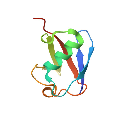 | |
UniProt & NIH Common Fund Data Resources | |||||
PHAROS: P0CG48 GTEx: ENSG00000150991 | |||||
Entity Groups | |||||
| Sequence Clusters | 30% Identity50% Identity70% Identity90% Identity95% Identity100% Identity | ||||
| UniProt Group | P0CG48 | ||||
Sequence AnnotationsExpand | |||||
Reference Sequence | |||||
| Ligands 1 Unique | |||||
|---|---|---|---|---|---|
| ID | Chains | Name / Formula / InChI Key | 2D Diagram | 3D Interactions | |
| ZN Download:Ideal Coordinates CCD File | D [auth A], E [auth A], F [auth A], G [auth B], H [auth B] | ZINC ION Zn PTFCDOFLOPIGGS-UHFFFAOYSA-N |  | ||
| Length ( Å ) | Angle ( ˚ ) |
|---|---|
| a = 43.786 | α = 90 |
| b = 49.735 | β = 90 |
| c = 93.168 | γ = 90 |
| Software Name | Purpose |
|---|---|
| CNS | refinement |
| CrystalClear | data collection |
| HKL-2000 | data reduction |
| HKL-2000 | data scaling |
| MOLREP | phasing |


