


Crystal Structure of Muconate Cycloisomerase from Thermotoga maritima MSB8
Padmanabhan, B., Bessho, Y., Yokoyama, S.To be published.
Experimental Data Snapshot
wwPDB Validation 3D Report Full Report

Entity ID: 1 | |||||
|---|---|---|---|---|---|
| Molecule | Chains | Sequence Length | Organism | Details | Image |
| Muconate cycloisomerase | 345 | Thermotoga maritima MSB8 | Mutation(s): 0 EC: 5.5.1.1 (PDB Primary Data), 5.1.1.20 (UniProt) | 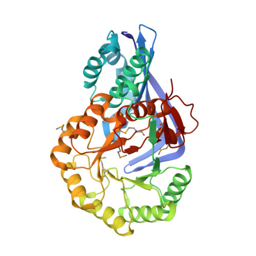 | |
UniProt | |||||
Entity Groups | |||||
| Sequence Clusters | 30% Identity50% Identity70% Identity90% Identity95% Identity100% Identity | ||||
| UniProt Group | Q9WXM1 | ||||
Sequence AnnotationsExpand | |||||
Reference Sequence | |||||
| Ligands 3 Unique | |||||
|---|---|---|---|---|---|
| ID | Chains | Name / Formula / InChI Key | 2D Diagram | 3D Interactions | |
| 1PE Download:Ideal Coordinates CCD File | F [auth A] G [auth A] H [auth A] L [auth B] M [auth B] | PENTAETHYLENE GLYCOL C10 H22 O6 JLFNLZLINWHATN-UHFFFAOYSA-N |  | ||
| PEG Download:Ideal Coordinates CCD File | I [auth A], J [auth A], N [auth B], W [auth D] | DI(HYDROXYETHYL)ETHER C4 H10 O3 MTHSVFCYNBDYFN-UHFFFAOYSA-N |  | ||
| MN Download:Ideal Coordinates CCD File | E [auth A], K [auth B], O [auth C], S [auth D] | MANGANESE (II) ION Mn WAEMQWOKJMHJLA-UHFFFAOYSA-N |  | ||
| Modified Residues 1 Unique | |||||
|---|---|---|---|---|---|
| ID | Chains | Type | Formula | 2D Diagram | Parent |
| MSE Query on MSE | A, B, C, D | L-PEPTIDE LINKING | C5 H11 N O2 Se |  | MET |
| Length ( Å ) | Angle ( ˚ ) |
|---|---|
| a = 112.77 | α = 90 |
| b = 112.77 | β = 90 |
| c = 122.526 | γ = 90 |
| Software Name | Purpose |
|---|---|
| REFMAC | refinement |
| ADSC | data collection |
| HKL-2000 | data reduction |
| SCALEPACK | data scaling |
| SOLVE | phasing |


