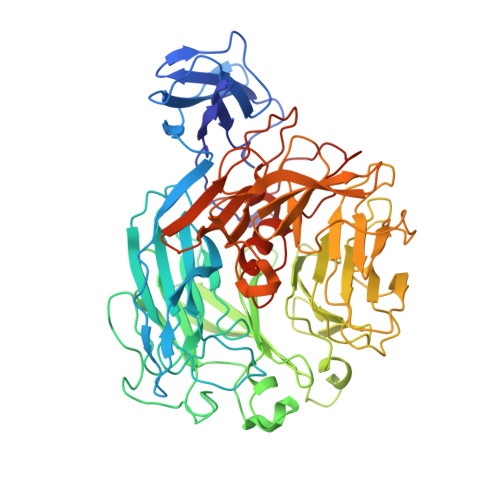
2Z8R

Crystal structure of rhamnogalacturonan lyase YesW at 1.40 A resolution


Experimental Data Snapshot

  • Method: X-RAY DIFFRACTION
  • Resolution: 1.40 Å
  • R-Value Free: 0.182 
  • R-Value Work: 0.164 
  • R-Value Observed: 0.164 

wwPDB Validation   3D Report Full Report


This is version 1.1 of the entry. See complete history


Literature

A Novel Structural Fold in Polysaccharide Lyases: BACILLUS SUBTILIS FAMILY 11 RHAMNOGALACTURONAN LYASE YesW WITH AN EIGHT-BLADED -PROPELLER

Ochiai, A.Itoh, T.Maruyama, Y.Kawamata, A.Mikami, B.Hashimoto, W.Murata, K.

(2007) J Biol Chem 282: 37134-37145

  • DOI: https://doi.org/10.1074/jbc.M704663200
  • Primary Citation of Related Structures:  
    2Z8R, 2Z8S

  • PubMed Abstract: 
  • Rhamnogalacturonan (RG) lyase produced by plant pathogenic and saprophytic microbes plays an important role in degrading plant cell walls. An extracellular RG lyase YesW from saprophytic Bacillus subtilis is a member of polysaccharide lyase family 11 and cleaves glycoside bonds in polygalacturonan as well as RG type-I through a beta-elimination reaction ...

    Rhamnogalacturonan (RG) lyase produced by plant pathogenic and saprophytic microbes plays an important role in degrading plant cell walls. An extracellular RG lyase YesW from saprophytic Bacillus subtilis is a member of polysaccharide lyase family 11 and cleaves glycoside bonds in polygalacturonan as well as RG type-I through a beta-elimination reaction. Crystal structures of YesW and its complex with galacturonan disaccharide, a reaction product analogue, were determined at 1.4 and 2.5 A resolutions with final R-factors of 16.4% and 16.6%, respectively. The enzyme is composed of an eight-bladed beta-propeller with a deep cleft in the center as a basic scaffold, and its structural fold has not been seen in polysaccharide lyases analyzed thus far. Structural analysis of the disaccharide-bound YesW and a site-directed mutagenesis study suggested that Arg-452 and Lys-535 stabilize the carboxyl group of the acidic polysaccharide molecule and Tyr-595 makes a stack interaction with the sugar pyranose ring. In addition to amino acid residues binding to the disaccharide, one calcium ion, which is coordinated by Asp-401, Glu-422, His-363, and His-399, may mediate the enzyme activity. This is, to our knowledge, the first report of a new structural category with a beta-propeller fold in polysaccharide lyases and provides structural insights into substrate binding by RG lyase.


    Organizational Affiliation

    Laboratory of Basic and Applied Molecular Biotechnology, Graduate School of Agriculture, Kyoto University, Japan.


Macromolecules
Find similar proteins by:  (by identity cutoff)  |  3D Structure
Entity ID: 1
MoleculeChainsSequence LengthOrganismDetailsImage
YesW proteinA, B591Bacillus subtilisMutation(s): 0 
Gene Names: yesW
EC: 4.2.2 (PDB Primary Data), 4.2.2.23 (UniProt)
UniProt
Find proteins for O31526 (Bacillus subtilis (strain 168))
Explore O31526 
Go to UniProtKB:  O31526
Entity Groups  
Sequence Clusters30% Identity50% Identity70% Identity90% Identity95% Identity100% Identity
UniProt GroupO31526
Protein Feature View
Expand
  • Reference Sequence
Small Molecules
Ligands 2 Unique
IDChainsName / Formula / InChI Key2D Diagram3D Interactions
MPD
Query on MPD

Download Ideal Coordinates CCD File 
CA [auth B],
DA [auth B],
EA [auth B],
FA [auth B],
M [auth A],
CA [auth B],
DA [auth B],
EA [auth B],
FA [auth B],
M [auth A],
N [auth A],
O [auth A],
P [auth A],
Q [auth A],
R [auth A]
(4S)-2-METHYL-2,4-PENTANEDIOL
C6 H14 O2
SVTBMSDMJJWYQN-YFKPBYRVSA-N
 Ligand Interaction
CA
Query on CA

Download Ideal Coordinates CCD File 
AA [auth B],
BA [auth B],
C [auth A],
D [auth A],
E [auth A],
AA [auth B],
BA [auth B],
C [auth A],
D [auth A],
E [auth A],
F [auth A],
G [auth A],
H [auth A],
I [auth A],
J [auth A],
K [auth A],
L [auth A],
S [auth B],
T [auth B],
U [auth B],
V [auth B],
W [auth B],
X [auth B],
Y [auth B],
Z [auth B]
CALCIUM ION
Ca
BHPQYMZQTOCNFJ-UHFFFAOYSA-N
 Ligand Interaction
Experimental Data & Validation

Experimental Data

  • Method: X-RAY DIFFRACTION
  • Resolution: 1.40 Å
  • R-Value Free: 0.182 
  • R-Value Work: 0.164 
  • R-Value Observed: 0.164 
  • Space Group: P 1 21 1
Unit Cell:
Length ( Å )Angle ( ˚ )
a = 56.897α = 90
b = 106.153β = 95.11
c = 101.041γ = 90
Software Package:
Software NamePurpose
REFMACrefinement
HKL-2000data collection
HKL-2000data reduction
HKL-2000data scaling
SOLVEphasing

Structure Validation

View Full Validation Report


Entry History 

Deposition Data

Revision History  (Full details and data files)

  • Version 1.0: 2007-10-16
    Type: Initial release
  • Version 1.1: 2011-07-13
    Changes: Version format compliance