


Crystal structure of H2O2-treated Cu,Zn-superoxide dismutase
Ito, S., Ishii, T., Uchida, K.To be published.
Experimental Data Snapshot
Starting Model: experimental
View more details
wwPDB Validation 3D Report Full Report

Entity ID: 1 | |||||
|---|---|---|---|---|---|
| Molecule | Chains | Sequence Length | Organism | Details | Image |
| Superoxide dismutase [Cu-Zn] | 151 | Bos taurus | Mutation(s): 0 EC: 1.15.1.1 (PDB Primary Data), 1.8 (UniProt) | 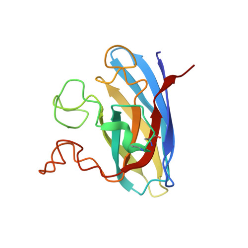 | |
UniProt | |||||
Entity Groups | |||||
| Sequence Clusters | 30% Identity50% Identity70% Identity90% Identity95% Identity100% Identity | ||||
| UniProt Group | P00442 | ||||
Sequence AnnotationsExpand | |||||
Reference Sequence | |||||
| Ligands 2 Unique | |||||
|---|---|---|---|---|---|
| ID | Chains | Name / Formula / InChI Key | 2D Diagram | 3D Interactions | |
| ZN Download:Ideal Coordinates CCD File | C [auth A], E [auth B] | ZINC ION Zn PTFCDOFLOPIGGS-UHFFFAOYSA-N |  | ||
| CU Download:Ideal Coordinates CCD File | D [auth A], F [auth B] | COPPER (II) ION Cu JPVYNHNXODAKFH-UHFFFAOYSA-N |  | ||
| Length ( Å ) | Angle ( ˚ ) |
|---|---|
| a = 45.344 | α = 90 |
| b = 50.822 | β = 90 |
| c = 148.993 | γ = 90 |
| Software Name | Purpose |
|---|---|
| CNS | refinement |
| HKL-2000 | data reduction |
| HKL-2000 | data scaling |
| SOLVE | phasing |


