2Z6U | pdb_00002z6u

Ternary structure of the Glu119Ala M.HhaI, C5-Cytosine DNA methyltransferase, with unmodified DNA and AdoHcy


Experimental Data Snapshot

  • Method: X-RAY DIFFRACTION
  • Resolution: 2.72 Å
  • R-Value Free: 
    0.267 (Depositor) 
  • R-Value Work: 
    0.212 (Depositor), 0.218 (DCC) 

wwPDB Validation 3D Report Full Report

Validation slider image for 2Z6U

Ligand Structure Quality Assessment 


This is version 1.3 of the entry. See complete history

Literature

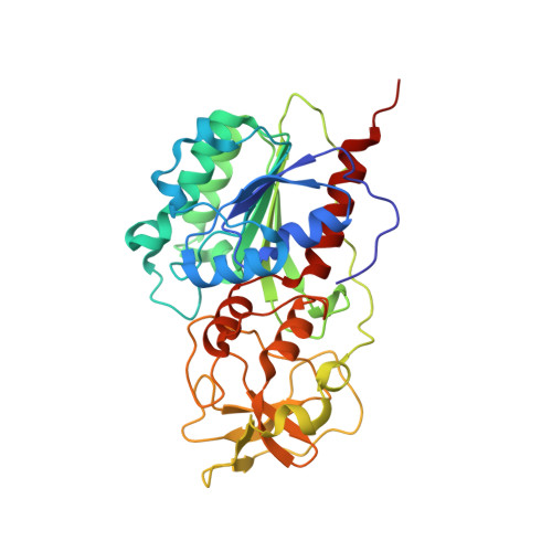
AdoMet-dependent Methyl-transfer: Glu(119) Is Essential for DNA C5-Cytosine Methyltransferase M.HhaI

Shieh, F.K.Reich, N.O.

(2007) J Mol Biology 373: 1157-1168

  • DOI: https://doi.org/10.1016/j.jmb.2007.08.009
  • Primary Citation Related Structures: 
    2Z6U, 2ZCJ

  • PubMed Abstract: 

    The role of Glu119 in S-adenosyl-L-methionine-dependent DNA methyltransferase M.HhaI-catalyzed DNA methylation was studied. Glu119 belongs to the highly conserved Glu/Asn/Val motif found in all DNA C5-cytosine methyltransferases, and its importance for M.HhaI function remains untested. We show that formation of the covalent intermediate between Cys81 and the target cytosine requires Glu119, since conversion to Ala, Asp or Gln lowers the rate of methyl transfer 10(2)-10(6) fold. Further, unlike the wild-type M.HhaI, these mutants are not trapped by the substrate in which the target cytosine is replaced with the mechanism-based inhibitor 5-fluorocytosine. The DNA binding affinity for the Glu119Asp mutant is decreased 10(3)-fold. Thus, the ability of the enzyme to stabilize the extrahelical cytosine is coupled directly to tight DNA binding. The structures of the ternary protein/DNA/AdoHcy complexes for both the Glu119Ala and Glu119Gln mutants (2.70 A and 2.75 A, respectively) show that the flipped base is positioned nearly identically with that observed in the wild-type M.HhaI complex. A single water molecule in the Glu119Ala structure between Ala119 and the extrahelical cytosine N3 is lacking in the Glu119Gln and wild-type M.HhaI structures, and most likely accounts for this mutant's partial activity. Glu119 has essential roles in activating the target cytosine for nucleophilic attack and contributes to tight DNA binding.


  • Organizational Affiliation
    • Department of Chemistry and Biochemistry, University of California, Santa Barbara, CA 93106-9510, USA.

Macromolecule Content 

  • Total Structure Weight: 45 kDa 
  • Atom Count: 3,229 
  • Modeled Residue Count: 352 
  • Deposited Residue Count: 352 
  • Unique protein chains: 1
  • Unique nucleic acid chains: 2

Macromolecules


Find similar proteins by:|  3D Structure
Entity ID: 3
MoleculeChains  Sequence LengthOrganismDetailsImage
Modification methylase HhaIC [auth A]327Haemophilus parahaemolyticusMutation(s): 1 
Gene Names: hhaIM
EC: 2.1.1.37
UniProt
Find proteins for P05102 (Haemophilus parahaemolyticus)
Explore P05102 
Go to UniProtKB:  P05102
Entity Groups
Sequence Clusters30% Identity50% Identity70% Identity90% Identity95% Identity100% Identity
UniProt GroupP05102
Sequence Annotations
Expand
Reference Sequence
Find similar nucleic acids by:  Sequence
Entity ID: 1
MoleculeChains LengthOrganismImage
DNA (5'-D(*DGP*DAP*DTP*DAP*DGP*DCP*DGP*DCP*DTP*DAP*DTP*DC)-3')A [auth C]12N/A
Sequence Annotations
Expand
Reference Sequence
Find similar nucleic acids by:  Sequence
Entity ID: 2
MoleculeChains LengthOrganismImage
DNA (5'-D(*DTP*DGP*DAP*DTP*DAP*DGP*DCP*DGP*DCP*DTP*DAP*DTP*DC)-3')B [auth D]13N/A
Sequence Annotations
Expand
Reference Sequence

Small Molecules

Ligands 1 Unique
IDChains Name / Formula / InChI Key2D Diagram3D Interactions
SAH

Query on SAH


Download:Ideal Coordinates CCD File
D [auth A]S-ADENOSYL-L-HOMOCYSTEINE
C14 H20 N6 O5 S
ZJUKTBDSGOFHSH-WFMPWKQPSA-N

Experimental Data & Validation

Experimental Data

  • Method: X-RAY DIFFRACTION
  • Resolution: 2.72 Å
  • R-Value Free:  0.267 (Depositor) 
  • R-Value Work:  0.212 (Depositor), 0.218 (DCC) 
Space Group: H 3 2
Unit Cell:
Length ( Å )Angle ( ˚ )
a = 98.41α = 90
b = 98.41β = 90
c = 322.31γ = 120
Software Package:
Software NamePurpose
HKL-2000data collection
CNSrefinement
HKL-2000data reduction
HKL-2000data scaling
CNSphasing

Structure Validation

View Full Validation Report


Ligand Structure Quality Assessment 


Entry History 

Deposition Data

  • Released Date: 2007-11-20 
  • Deposition Author(s): Shieh, F.K.

Revision History  (Full details and data files)

  • Version 1.0: 2007-11-20
    Type: Initial release
  • Version 1.1: 2011-07-13
    Changes: Version format compliance
  • Version 1.2: 2021-11-10
    Changes: Database references, Derived calculations
  • Version 1.3: 2024-05-29
    Changes: Data collection