2Z3U | pdb_00002z3u

Crystal Structure of Chromopyrrolic Acid Bound Cytochrome P450 StaP (CYP245A1)


Experimental Data Snapshot

  • Method: X-RAY DIFFRACTION
  • Resolution: 2.40 Å
  • R-Value Free: 
    0.238 (Depositor), 0.232 (DCC) 
  • R-Value Work: 
    0.191 (Depositor), 0.184 (DCC) 

Starting Model: experimental
View more details

wwPDB Validation 3D Report Full Report

Validation slider image for 2Z3U

Ligand Structure Quality Assessment 


This is version 1.3 of the entry. See complete history

Literature

Crystal structures and catalytic mechanism of cytochrome P450 StaP that produces the indolocarbazole skeleton

Makino, M.Sugimoto, H.Shiro, Y.Asamizu, S.Onaka, H.Nagano, S.

(2007) Proc Natl Acad Sci U S A 104: 11591-11596

  • DOI: https://doi.org/10.1073/pnas.0702946104
  • Primary Citation Related Structures: 
    2Z3T, 2Z3U

  • PubMed Abstract: 

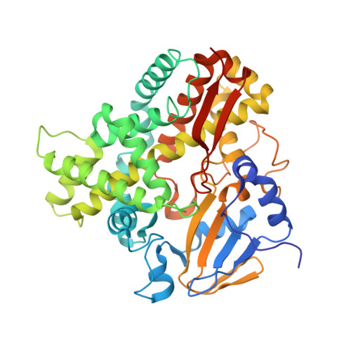
    Staurosporine isolated from Streptomyces sp. TP-A0274 is a member of the family of indolocarbazole alkaloids that exhibit strong antitumor activity. A key step in staurosporine biosynthesis is the formation of the indolocarbazole core by intramolecular C-C bond formation and oxidative decarboxylation of chromopyrrolic acid (CPA) catalyzed by cytochrome P450 StaP (StaP, CYP245A1). In this study, we report x-ray crystal structures of CPA-bound and -free forms of StaP. Upon substrate binding, StaP adopts a more ordered conformation, and conformational rearrangements of residues in the active site are also observed. Hydrogen-bonding interactions of two carboxyl groups and T-shaped pi-pi interactions with indole rings hold the substrate in the substrate-binding cavity with a conformation perpendicular to the heme plane. Based on the crystal structure of StaP-CPA complex, we propose that C-C bond formation occurs through an indole cation radical intermediate that is equivalent to cytochrome c peroxidase compound I [Sivaraja M, Goodin DB, Smith M, Hoffman BM (1989) Science 245:738-740]. The subsequent oxidative decarboxylation reaction is also discussed based on the crystal structure. Our crystallographic study shows the first crystal structures of enzymes involved in formation of the indolocarbazole core and provides valuable insights into the process of staurosporine biosynthesis, combinatorial biosynthesis of indolocarbazoles, and the diversity of cytochrome P450 chemistry.


  • Organizational Affiliation
    • Biometal Science Laboratory, RIKEN SPring-8 Center, Harima Institute, Hyogo 679-5148, Japan.

Macromolecule Content 

  • Total Structure Weight: 49.28 kDa 
  • Atom Count: 3,552 
  • Modeled Residue Count: 411 
  • Deposited Residue Count: 425 
  • Unique protein chains: 1

Macromolecules

Find similar proteins by:|  3D Structure
Entity ID: 1
MoleculeChains  Sequence LengthOrganismDetailsImage
Cytochrome P450425Streptomyces sp. TP-A0274Mutation(s): 0 
Gene Names: staP
EC: 1
UniProt
Find proteins for Q83WG3 (Streptomyces sp. TP-A0274)
Explore Q83WG3 
Go to UniProtKB:  Q83WG3
Entity Groups
Sequence Clusters30% Identity50% Identity70% Identity90% Identity95% Identity100% Identity
UniProt GroupQ83WG3
Sequence Annotations
Expand
Reference Sequence

Small Molecules

Ligands 3 Unique
IDChains Name / Formula / InChI Key2D Diagram3D Interactions
HEM

Query on HEM


Download:Ideal Coordinates CCD File
B [auth A]PROTOPORPHYRIN IX CONTAINING FE
C34 H32 Fe N4 O4
KABFMIBPWCXCRK-RGGAHWMASA-L
CRR

Query on CRR


Download:Ideal Coordinates CCD File
C [auth A],
D [auth A],
E [auth A]
3,4-DI-1H-INDOL-3-YL-1H-PYRROLE-2,5-DICARBOXYLIC ACID
C22 H15 N3 O4
FZDVNXHYGMEEDT-UHFFFAOYSA-N
EDO

Query on EDO


Download:Ideal Coordinates CCD File
F [auth A],
G [auth A],
H [auth A],
I [auth A]
1,2-ETHANEDIOL
C2 H6 O2
LYCAIKOWRPUZTN-UHFFFAOYSA-N

Experimental Data & Validation

Experimental Data

  • Method: X-RAY DIFFRACTION
  • Resolution: 2.40 Å
  • R-Value Free:  0.238 (Depositor), 0.232 (DCC) 
  • R-Value Work:  0.191 (Depositor), 0.184 (DCC) 
Space Group: P 21 21 21
Unit Cell:
Length ( Å )Angle ( ˚ )
a = 44.627α = 90
b = 66.61β = 90
c = 136.813γ = 90
Software Package:
Software NamePurpose
MOLREPphasing
CNSrefinement
PDB_EXTRACTdata extraction
BSSdata collection
HKL-2000data reduction
SCALEPACKdata scaling

Structure Validation

View Full Validation Report


Ligand Structure Quality Assessment 


Entry History 

Deposition Data

Revision History  (Full details and data files)

  • Version 1.0: 2007-07-03
    Type: Initial release
  • Version 1.1: 2008-04-30
    Changes: Version format compliance
  • Version 1.2: 2011-07-13
    Changes: Version format compliance
  • Version 1.3: 2023-11-01
    Changes: Data collection, Database references, Derived calculations, Refinement description