2Z35 | pdb_00002z35

Crystal structure of immune receptor


Experimental Data Snapshot

  • Method: X-RAY DIFFRACTION
  • Resolution: 2.20 Å
  • R-Value Free: 
    0.285 (Depositor), 0.264 (DCC) 
  • R-Value Work: 
    0.263 (Depositor), 0.271 (DCC) 

wwPDB Validation 3D Report Full Report

Validation slider image for 2Z35

This is version 1.2 of the entry. See complete history

Literature

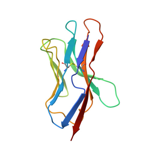
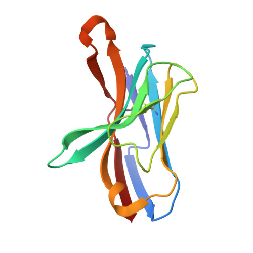
Structural evidence for a germline-encoded T cell receptor-major histocompatibility complex interaction 'codon'

Feng, D.Bond, C.J.Ely, L.K.Maynard, J.Garcia, K.C.

(2007) Nat Immunol 8: 975-983

  • DOI: https://doi.org/10.1038/ni1502
  • Primary Citation Related Structures: 
    2PXY, 2Z31, 2Z35

  • PubMed Abstract: 

    All complexes of T cell receptors (TCRs) bound to peptide-major histocompatibility complex (pMHC) molecules assume a stereotyped binding 'polarity', despite wide variations in TCR-pMHC docking angles. However, existing TCR-pMHC crystal structures have failed to show broadly conserved pairwise interaction motifs. Here we determined the crystal structures of two TCRs encoded by the variable beta-chain 8.2 (V(beta)8.2), each bound to the MHC class II molecule I-A(u), and did energetic mapping of V(alpha) and V(beta) contacts with I-A(u). Together with two previously solved structures of V(beta)8.2-containing TCR-MHC complexes, we found four TCR-I-A complexes with structurally superimposable interactions between the V(beta) loops and the I-A alpha-helix. This examination of a narrow 'slice' of the TCR-MHC repertoire demonstrates what is probably one of many germline-derived TCR-MHC interaction 'codons'.


  • Organizational Affiliation
    • Howard Hughes Medical Institute, Department of Molecular & Cellular Physiology, Stanford University School of Medicine, Stanford, California 94305, USA.

Macromolecule Content 

  • Total Structure Weight: 24.48 kDa 
  • Atom Count: 1,751 
  • Modeled Residue Count: 223 
  • Deposited Residue Count: 223 
  • Unique protein chains: 2

Macromolecules

Find similar proteins by:|  3D Structure
Entity ID: 1
MoleculeChains  Sequence LengthOrganismDetailsImage
T-cell receptor alpha-chain112Mus musculusMutation(s): 0 
UniProt
Find proteins for Q5R1F5 (Mus musculus)
Explore Q5R1F5 
Go to UniProtKB:  Q5R1F5
Entity Groups
Sequence Clusters30% Identity50% Identity70% Identity90% Identity95% Identity100% Identity
UniProt GroupQ5R1F5
Sequence Annotations
Expand
Reference Sequence
Find similar proteins by:|  3D Structure
Entity ID: 2
MoleculeChains  Sequence LengthOrganismDetailsImage
T-cell receptor beta-chain111Mus musculusMutation(s): 0 
UniProt
Find proteins for A2NTY6 (Mus musculus)
Explore A2NTY6 
Go to UniProtKB:  A2NTY6
Entity Groups
Sequence Clusters30% Identity50% Identity70% Identity90% Identity95% Identity100% Identity
UniProt GroupA2NTY6
Sequence Annotations
Expand
Reference Sequence

Experimental Data & Validation

Experimental Data

  • Method: X-RAY DIFFRACTION
  • Resolution: 2.20 Å
  • R-Value Free:  0.285 (Depositor), 0.264 (DCC) 
  • R-Value Work:  0.263 (Depositor), 0.271 (DCC) 
Space Group: I 2 2 2
Unit Cell:
Length ( Å )Angle ( ˚ )
a = 67.994α = 90
b = 73.802β = 90
c = 115.374γ = 90
Software Package:
Software NamePurpose
HKL-2000data collection
AMoREphasing
CNSrefinement
HKL-2000data reduction
HKL-2000data scaling

Structure Validation

View Full Validation Report


Entry History 

Deposition Data

Revision History  (Full details and data files)

  • Version 1.0: 2007-10-09
    Type: Initial release
  • Version 1.1: 2011-07-13
    Changes: Version format compliance
  • Version 1.2: 2024-10-30
    Changes: Data collection, Database references, Structure summary