2Z23 | pdb_00002z23

Crystal structure of Y.pestis oligo peptide binding protein OppA with tri-lysine ligand


Experimental Data Snapshot

  • Method: X-RAY DIFFRACTION
  • Resolution: 2.00 Å
  • R-Value Free: 
    0.239 (Depositor), 0.242 (DCC) 
  • R-Value Work: 
    0.210 (Depositor), 0.214 (DCC) 
  • R-Value Observed: 
    0.210 (Depositor) 

wwPDB Validation 3D Report Full Report

Validation slider image for 2Z23

This is version 1.2 of the entry. See complete history

Literature

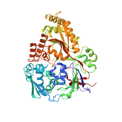
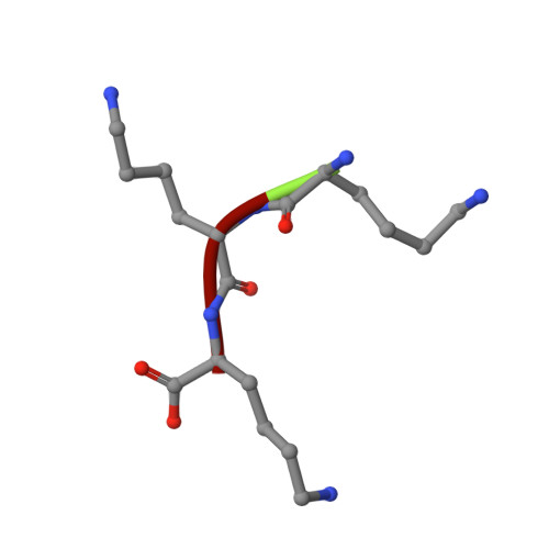
Structures of OppA and PstS from Yersinia pestis indicate variability of interactions with transmembrane domains.

Tanabe, M.Mirza, O.Bertrand, T.Atkins, H.S.Titball, R.W.Iwata, S.Brown, K.A.Byrne, B.

(2007) Acta Crystallogr D Biol Crystallogr 63: 1185-1193

  • DOI: https://doi.org/10.1107/S0907444907048299
  • Primary Citation Related Structures: 
    2Z22, 2Z23

  • PubMed Abstract: 

    Bacterial ATP-binding cassette (ABC) transport systems couple ATP hydrolysis with the uptake and efflux of a wide range of substances across bacterial membranes. These systems are comprised of transmembrane domains, nucleotide binding domains and, in the case of uptake systems, periplasmic binding proteins responsible for binding and presentation of substrate to the transmembrane domains. In pathogenic bacteria, ABC systems are known to play roles in virulence and pathogenicity and the surface localization of some components has made them attractive targets for both vaccine and anti-infective development. Here, the crystallization of five proteins (OppA, PstS, PiuA, YrbD and CysP) from Yersinia pestis, the causative agent of plague, are reported that diffracted to resolution limits ranging from 1.6 to 5 A. The first crystal structures of ABC system components from Y. pestis, OppA and PstS, are also reported here as complexes with their substrates. Comparisons of these two structures with known structures of related proteins suggest that these proteins possess versatility in substrate recognition and variations in protein-protein interactions with their cognate transmembrane domains.


  • Organizational Affiliation
    • Membrane Protein Crystallography, Division of Molecular Biosciences, Imperial College London, London SW7 2AZ, England.

Macromolecule Content 

  • Total Structure Weight: 59.26 kDa 
  • Atom Count: 4,570 
  • Modeled Residue Count: 520 
  • Deposited Residue Count: 520 
  • Unique protein chains: 2

Macromolecules

Find similar proteins by:|  3D Structure
Entity ID: 1
MoleculeChains  Sequence LengthOrganismDetailsImage
Periplasmic oligopeptide-binding protein517Yersinia pestis CO92Mutation(s): 0 
UniProt
Find proteins for A0A0H2W5P5 (Yersinia pestis)
Explore A0A0H2W5P5 
Go to UniProtKB:  A0A0H2W5P5
Entity Groups
Sequence Clusters30% Identity50% Identity70% Identity90% Identity95% Identity100% Identity
UniProt GroupA0A0H2W5P5
Sequence Annotations
Expand
Reference Sequence
Find similar proteins by:  Sequence   |   3D Structure  
Entity ID: 2
MoleculeChains  Sequence LengthOrganismDetailsImage
peptide (LYS)(LYS)(LYS)3N/AMutation(s): 0 

Experimental Data & Validation

Experimental Data

  • Method: X-RAY DIFFRACTION
  • Resolution: 2.00 Å
  • R-Value Free:  0.239 (Depositor), 0.242 (DCC) 
  • R-Value Work:  0.210 (Depositor), 0.214 (DCC) 
  • R-Value Observed: 0.210 (Depositor) 
Space Group: P 21 21 21
Unit Cell:
Length ( Å )Angle ( ˚ )
a = 71.827α = 90
b = 90.637β = 90
c = 93.702γ = 90
Software Package:
Software NamePurpose
PHASERphasing
CNSrefinement
ADSCdata collection
DENZOdata reduction
SCALEPACKdata scaling

Structure Validation

View Full Validation Report


Entry History 

Deposition Data

Revision History  (Full details and data files)

  • Version 1.0: 2007-10-30
    Type: Initial release
  • Version 1.1: 2011-07-13
    Changes: Source and taxonomy, Version format compliance
  • Version 1.2: 2024-11-06
    Changes: Data collection, Database references, Structure summary