


Crystal Structure of Ttha1563 from Thermus thermophilus HB8
Niwa, H., Shimada, A., Matsunaga, E., Kuramitsu, S., Yokoyama, S.To be published.
Experimental Data Snapshot
Starting Model: experimental
View more details
wwPDB Validation 3D Report Full Report

Macromolecule Content
Entity ID: 1 | |||||
|---|---|---|---|---|---|
| Molecule | Chains | Sequence Length | Organism | Details | Image |
| (Neo)pullulanase | 475 | Thermus thermophilus HB8 | Mutation(s): 0 EC: 3.2.1.41 | 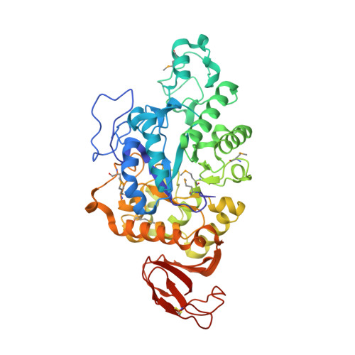 | |
UniProt | |||||
Entity Groups | |||||
| Sequence Clusters | 30% Identity50% Identity70% Identity90% Identity95% Identity100% Identity | ||||
| UniProt Group | Q5SI17 | ||||
Sequence AnnotationsExpand | |||||
Reference Sequence | |||||
Entity ID: 2 | |||||
|---|---|---|---|---|---|
| Molecule | Chains | Length | 2D Diagram | Glycosylation | D Interactions |
| Cycloheptakis-(1-4)-(alpha-D-glucopyranose) | E, G, I, K | 7 |  | N/A | |
Glycosylation Resources | |||||
GlyTouCan: G01435GL GlyCosmos: G01435GL | |||||
| Ligands 1 Unique | |||||
|---|---|---|---|---|---|
| ID | Chains | Name / Formula / InChI Key | 2D Diagram | 3D Interactions | |
| PO4 Download:Ideal Coordinates CCD File | M [auth D] | PHOSPHATE ION O4 P NBIIXXVUZAFLBC-UHFFFAOYSA-K |  | ||
| Modified Residues 1 Unique | |||||
|---|---|---|---|---|---|
| ID | Chains | Type | Formula | 2D Diagram | Parent |
| MSE Query on MSE | A, B, C, D | L-PEPTIDE LINKING | C5 H11 N O2 Se |  | MET |
Entity ID: 2 | |||||
|---|---|---|---|---|---|
| ID | Chains | Name | Type/Class | 2D Diagram | 3D Interactions |
| PRD_900012 Query on PRD_900012 | E, G, I, K | beta-cyclodextrin | Oligosaccharide / Drug delivery |  |
Entity ID: 3 | |||||
|---|---|---|---|---|---|
| ID | Chains | Name | Type/Class | 2D Diagram | 3D Interactions |
| PRD_900009 Query on PRD_900009 | F, H, J, L | alpha-maltotriose | Oligosaccharide / Nutrient |  |
| Length ( Å ) | Angle ( ˚ ) |
|---|---|
| a = 67.144 | α = 90 |
| b = 95.897 | β = 94.4 |
| c = 140.598 | γ = 90 |
| Software Name | Purpose |
|---|---|
| CNS | refinement |
| HKL-2000 | data collection |
| HKL-2000 | data reduction |
| HKL-2000 | data scaling |
| PHASER | phasing |


