2YVC

Crystal structure of the Radixin FERM domain complexed with the NEP cytoplasmic tail


Experimental Data Snapshot

  • Method: X-RAY DIFFRACTION
  • Resolution: 3.20 Å
  • R-Value Free: 0.267 
  • R-Value Work: 0.233 
  • R-Value Observed: 0.234 

wwPDB Validation   3D Report Full Report


This is version 1.3 of the entry. See complete history


Literature

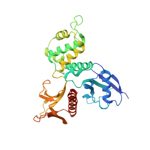
Structural basis for type II membrane protein binding by ERM proteins revealed by the radixin-neutral endopeptidase 24.11 (NEP) complex

Terawaki, S.Kitano, K.Hakoshima, T.

(2007) J Biol Chem 282: 19854-19862

  • DOI: https://doi.org/10.1074/jbc.M609232200
  • Primary Citation of Related Structures:  
    2YVC

  • PubMed Abstract: 

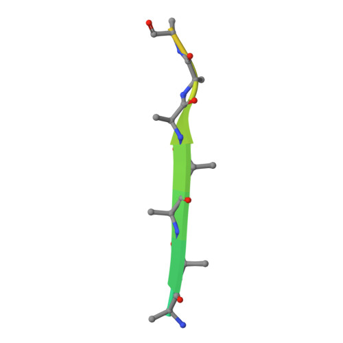
    ERM (Ezrin/Radixin/Moesin) proteins mediate formation of membrane-associated cytoskeletons by simultaneously binding actin filaments and the C-terminal cytoplasmic tails of adhesion molecules (type I membrane proteins). ERM proteins also bind neutral endopeptidase 24.11 (NEP), a type II membrane protein, even though the N-terminal cytoplasmic tail of NEP possesses the opposite peptide polarity to that of type I membrane proteins. Here, we determined the crystal structure of the radixin FERM (Four point one and ERM) domain complexed with the N-terminal NEP cytoplasmic peptide. In the FERM-NEP complex, the amphipathic region of the peptide forms a beta strand followed by a hairpin that bind to a shallow groove of FERM subdomain C. NEP binding is stabilized by beta-beta interactions and docking of the NEP hairpin into the hydrophobic pocket of subdomain C. Whereas the binding site of NEP on the FERM domain overlaps with the binding site of intercellular adhesion molecule (ICAM)-2, NEP lacks the Motif-1 sequence conserved in ICAM-2 and related adhesion molecules. The NEP hairpin, although lacking the typical inter-chain hydrogen bond but is stabilized by hydrogen bonds with the main chain and side chains of subdomain C, directs the C-terminal basic region of the NEP peptide away from the groove and toward the membrane. The overlap of the binding sites on subdomain C for NEP and Motif-1 adhesion molecules such as CD44 provides the structural basis for the suppression of cell adhesion through interaction between NEP and ERM proteins.


  • Organizational Affiliation

    Structural Biology Laboratory, Nara Institute of Science and Technology, and CREST, Japan Science and Technology Agency, 8916-5 Takayama, Ikoma, Nara 630-0192, Japan.


Macromolecules
Find similar proteins by:  (by identity cutoff)  |  3D Structure
Entity ID: 1
MoleculeChains Sequence LengthOrganismDetailsImage
Radixin
A, B, C
312Mus musculusMutation(s): 0 
UniProt & NIH Common Fund Data Resources
Find proteins for P26043 (Mus musculus)
Explore P26043 
Go to UniProtKB:  P26043
IMPC:  MGI:97887
Entity Groups  
Sequence Clusters30% Identity50% Identity70% Identity90% Identity95% Identity100% Identity
UniProt GroupP26043
Sequence Annotations
Expand
  • Reference Sequence

Find similar proteins by:  Sequence   |   3D Structure  

Entity ID: 2
MoleculeChains Sequence LengthOrganismDetailsImage
Neprilysin
D, E, F
22N/AMutation(s): 0 
UniProt & NIH Common Fund Data Resources
Find proteins for Q61391 (Mus musculus)
Explore Q61391 
Go to UniProtKB:  Q61391
IMPC:  MGI:97004
Entity Groups  
Sequence Clusters30% Identity50% Identity70% Identity90% Identity95% Identity100% Identity
UniProt GroupQ61391
Sequence Annotations
Expand
  • Reference Sequence
Experimental Data & Validation

Experimental Data

  • Method: X-RAY DIFFRACTION
  • Resolution: 3.20 Å
  • R-Value Free: 0.267 
  • R-Value Work: 0.233 
  • R-Value Observed: 0.234 
  • Space Group: P 21 21 21
Unit Cell:
Length ( Å )Angle ( ˚ )
a = 108.79α = 90
b = 116.838β = 90
c = 141.884γ = 90
Software Package:
Software NamePurpose
REFMACrefinement
HKL-2000data collection
HKL-2000data reduction
HKL-2000data scaling
MOLREPphasing

Structure Validation

View Full Validation Report


Entry History 

Deposition Data

Revision History  (Full details and data files)

  • Version 1.0: 2007-04-24
    Type: Initial release
  • Version 1.1: 2008-04-30
    Changes: Version format compliance
  • Version 1.2: 2011-07-13
    Changes: Version format compliance
  • Version 1.3: 2023-10-25
    Changes: Data collection, Database references, Refinement description