The interaction of DiaA and DnaA regulates the replication cycle in E. coli by directly promoting ATP DnaA-specific initiation complexes
Keyamura, K., Fujikawa, N., Ishida, T., Ozaki, S., Suetsugu, M., Fujimitsu, K., Kagawa, W., Yokoyama, S., Kurumizaka, H., Katayama, T.(2007) Genes Dev 21: 2083-2099
- PubMed: 17699754 Search on PubMedSearch on PubMed Central
- DOI: https://doi.org/10.1101/gad.1561207
- Primary Citation Related Structures:
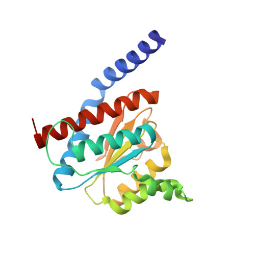
2YVA - PubMed Abstract:
Escherichia coli DiaA is a DnaA-binding protein that is required for the timely initiation of chromosomal replication during the cell cycle. In this study, we determined the crystal structure of DiaA at 1.8 A resolution. DiaA forms a homotetramer consisting of a symmetrical pair of homodimers. Mutational analysis revealed that the DnaA-binding activity and formation of homotetramers are required for the stimulation of initiation by DiaA. DiaA tetramers can bind multiple DnaA molecules simultaneously. DiaA stimulated the assembly of multiple DnaA molecules on oriC, conformational changes in ATP-DnaA-specific initiation complexes, and unwinding of oriC duplex DNA. The mutant DiaA proteins are defective in these stimulations. DiaA associated also with ADP-DnaA, and stimulated the assembly of inactive ADP-DnaA-oriC complexes. Specific residues in the putative phosphosugar-binding motif of DiaA were required for the stimulation of initiation and formation of ATP-DnaA-specific-oriC complexes. Our data indicate that DiaA regulates initiation by a novel mechanism, in which DiaA tetramers most likely bind to multiple DnaA molecules and stimulate the assembly of specific ATP-DnaA-oriC complexes. These results suggest an essential role for DiaA in the promotion of replication initiation in a cell cycle coordinated manner.
- Department of Molecular Biology, Graduate School of Pharmaceutical Sciences, Kyushu University, Fukuoka 812-8582, Japan.
Organizational Affiliation: