


Structure of the Epsilon-Lysine Oxidase from Marinomonas Mediterranea
Medrano, F.J., Sanchez-Amat, A., Romero, A.To be published.
Experimental Data Snapshot
wwPDB Validation 3D Report Full Report

Entity ID: 1 | |||||
|---|---|---|---|---|---|
| Molecule | Chains | Sequence Length | Organism | Details | Image |
| L-LYSINE 6-OXIDASE | 746 | Marinomonas mediterranea MMB-1 | Mutation(s): 0 EC: 1.4.3.20 | 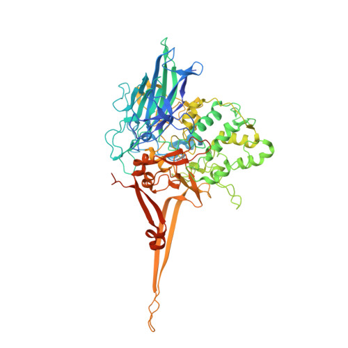 | |
UniProt | |||||
Entity Groups | |||||
| Sequence Clusters | 30% Identity50% Identity70% Identity90% Identity95% Identity100% Identity | ||||
| UniProt Group | F2JXJ3 | ||||
Sequence AnnotationsExpand | |||||
Reference Sequence | |||||
| Ligands 3 Unique | |||||
|---|---|---|---|---|---|
| ID | Chains | Name / Formula / InChI Key | 2D Diagram | 3D Interactions | |
| GOL Download:Ideal Coordinates CCD File | WB [auth B], XB [auth B] | GLYCEROL C3 H8 O3 PEDCQBHIVMGVHV-UHFFFAOYSA-N |  | ||
| EOH Download:Ideal Coordinates CCD File | AA [auth A] AB [auth B] AC [auth B] BA [auth A] BB [auth B] | ETHANOL C2 H6 O LFQSCWFLJHTTHZ-UHFFFAOYSA-N |  | ||
| NA Download:Ideal Coordinates CCD File | C [auth A], D [auth A], VA [auth B], WA [auth B] | SODIUM ION Na FKNQFGJONOIPTF-UHFFFAOYSA-N |  | ||
| Modified Residues 1 Unique | |||||
|---|---|---|---|---|---|
| ID | Chains | Type | Formula | 2D Diagram | Parent |
| TRQ Query on TRQ | A, B | L-PEPTIDE LINKING | C11 H10 N2 O4 |  | TRP |
| Length ( Å ) | Angle ( ˚ ) |
|---|---|
| a = 70.68 | α = 90 |
| b = 127.883 | β = 107.18 |
| c = 106.897 | γ = 90 |
| Software Name | Purpose |
|---|---|
| BUSTER | refinement |
| XDS | data reduction |
| XDS | data scaling |
| SHARP | phasing |


