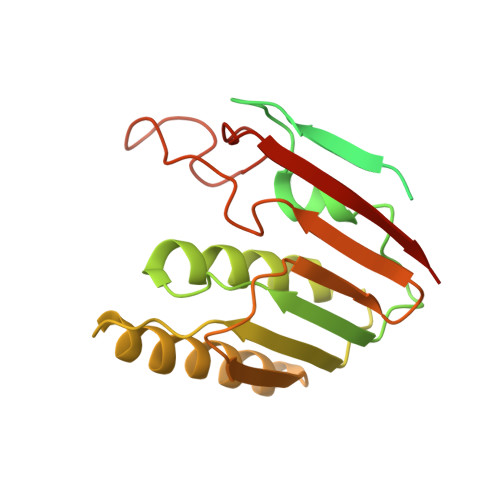
2YMB | pdb_00002ymb

Structures of MITD1


Experimental Data Snapshot

  • Method: X-RAY DIFFRACTION
  • Resolution: 3.40 Å
  • R-Value Free: 
    0.256 (Depositor), 0.254 (DCC) 
  • R-Value Work: 
    0.230 (Depositor), 0.223 (DCC) 
  • R-Value Observed: 
    0.230 (Depositor) 

Starting Models: experimental
View more details

wwPDB Validation 3D Report Full Report

Validation slider image for 2YMB

This is version 1.2 of the entry. See complete history. 

Literature

Escrt-III Binding Protein Mitd1 is Involved in Cytokinesis and Has an Unanticipated Pld Fold that Binds Membranes.

Hadders, M.A., Agromayor, M., Obita, T., Perisic, O., Caballe, A., Kloc, M., Lamers, M.H., Williams, R.L., Martin-Serrano, J.

(2012) Proc Natl Acad Sci U S A 109: 17424

  • DOI: https://doi.org/10.1073/pnas.1206839109
  • Primary Citation Related Structures: 
    2YMB, 4A5X, 4A5Z

  • PubMed Abstract: 

    The endosomal sorting complexes required for transport (ESCRT) proteins have a critical function in abscission, the final separation of the daughter cells during cytokinesis. Here, we describe the structure and function of a previously uncharacterized ESCRT-III interacting protein, MIT-domain containing protein 1 (MITD1). Crystal structures of MITD1 reveal a dimer, with a microtubule-interacting and trafficking (MIT) domain at the N terminus and a unique, unanticipated phospholipase D-like (PLD) domain at the C terminus that binds membranes. We show that the MIT domain binds to a subset of ESCRT-III subunits and that this interaction mediates MITD1 recruitment to the midbody during cytokinesis. Depletion of MITD1 causes a distinct cytokinetic phenotype consistent with destabilization of the midbody and abscission failure. These results suggest a model whereby MITD1 coordinates the activity of ESCRT-III during abscission with earlier events in the final stages of cell division.


  • Organizational Affiliation: 
    • Laboratory of Molecular Biology, Medical Research Council, Cambridge CB2 0QH, United Kingdom.

Macromolecule Content 

  • Total Structure Weight: 124.93 kDa 
  • Atom Count: 6,629 
  • Modeled Residue Count: 794 
  • Deposited Residue Count: 1,056 
  • Unique protein chains: 2

Macromolecules

Find similar proteins by:|  3D Structure
Entity ID: 1
MoleculeChains  Sequence LengthOrganismDetailsImage
MIT DOMAIN-CONTAINING PROTEIN 1
A, B, C, D
257Homo sapiensMutation(s): 0 
UniProt & NIH Common Fund Data Resources
Find proteins for Q8WV92 (Homo sapiens)
Explore Q8WV92 
Go to UniProtKB:  Q8WV92
PHAROS:  Q8WV92
GTEx:  ENSG00000158411 
Entity Groups
Sequence Clusters30% Identity50% Identity70% Identity90% Identity95% Identity100% Identity
UniProt GroupQ8WV92
Sequence Annotations
Expand
Reference Sequence
Find similar proteins by:  Sequence   |   3D Structure  
Entity ID: 2
MoleculeChains  Sequence LengthOrganismDetailsImage
CHARGED MULTIVESICULAR BODY PROTEIN 1AE [auth F],
F [auth H]
14Homo sapiensMutation(s): 0 
UniProt & NIH Common Fund Data Resources
Find proteins for Q9HD42 (Homo sapiens)
Explore Q9HD42 
Go to UniProtKB:  Q9HD42
PHAROS:  Q9HD42
GTEx:  ENSG00000131165 
Entity Groups
Sequence Clusters30% Identity50% Identity70% Identity90% Identity95% Identity100% Identity
UniProt GroupQ9HD42
Sequence Annotations
Expand
Reference Sequence

Experimental Data & Validation

Experimental Data

  • Method: X-RAY DIFFRACTION
  • Resolution: 3.40 Å
  • R-Value Free:  0.256 (Depositor), 0.254 (DCC) 
  • R-Value Work:  0.230 (Depositor), 0.223 (DCC) 
  • R-Value Observed: 0.230 (Depositor) 
Space Group: I 2 3
Unit Cell:
Length ( Å )Angle ( ˚ )
a = 222.046α = 90
b = 222.046β = 90
c = 222.046γ = 90
Software Package:
Software NamePurpose
PHENIXrefinement
XDSdata reduction
SCALAdata scaling
PHASERphasing

Structure Validation

View Full Validation Report


Entry History 

Deposition Data

Revision History  (Full details and data files)

  • Version 1.0: 2012-10-24
    Type: Initial release
  • Version 1.1: 2012-11-07
    Changes: Database references
  • Version 1.2: 2023-12-20
    Changes: Data collection, Database references, Other, Refinement description