2YKG | pdb_00002ykg

Structural insights into RNA recognition by RIG-I


Experimental Data Snapshot

  • Method: X-RAY DIFFRACTION
  • Resolution: 2.50 Å
  • R-Value Free: 
    0.273 (Depositor), 0.239 (DCC) 
  • R-Value Work: 
    0.220 (Depositor) 
  • R-Value Observed: 
    0.223 (Depositor) 

wwPDB Validation 3D Report Full Report

Validation slider image for 2YKG

This is version 1.1 of the entry. See complete history

Literature

Structural Insights Into RNA Recognition by Rig-I.

Luo, D.Ding, S.C.Vela, A.Kohlway, A.Lindenbach, B.D.Pyle, A.M.

(2011) Cell 147: 409

  • DOI: https://doi.org/10.1016/j.cell.2011.09.023
  • Primary Citation Related Structures: 
    2YKG, 4BPB

  • PubMed Abstract: 

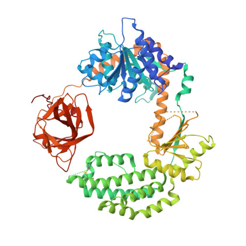
    Intracellular RIG-I-like receptors (RLRs, including RIG-I, MDA-5, and LGP2) recognize viral RNAs as pathogen-associated molecular patterns (PAMPs) and initiate an antiviral immune response. To understand the molecular basis of this process, we determined the crystal structure of RIG-I in complex with double-stranded RNA (dsRNA). The dsRNA is sheathed within a network of protein domains that include a conserved "helicase" domain (regions HEL1 and HEL2), a specialized insertion domain (HEL2i), and a C-terminal regulatory domain (CTD). A V-shaped pincer connects HEL2 and the CTD by gripping an α-helical shaft that extends from HEL1. In this way, the pincer coordinates functions of all the domains and couples RNA binding with ATP hydrolysis. RIG-I falls within the Dicer-RIG-I clade of the superfamily 2 helicases, and this structure reveals complex interplay between motor domains, accessory mechanical domains, and RNA that has implications for understanding the nanomechanical function of this protein family and other ATPases more broadly.


  • Organizational Affiliation
    • Department of Molecular, Cellular, and Developmental Biology, Yale University, New Haven, CT 06520, USA.

Macromolecule Content 

  • Total Structure Weight: 86.26 kDa 
  • Atom Count: 5,523 
  • Modeled Residue Count: 656 
  • Deposited Residue Count: 716 
  • Unique protein chains: 1
  • Unique nucleic acid chains: 1

Macromolecules


Find similar proteins by:|  3D Structure
Entity ID: 1
MoleculeChains  Sequence LengthOrganismDetailsImage
PROBABLE ATP-DEPENDENT RNA HELICASE DDX58696Homo sapiensMutation(s): 0 
EC: 3.6.4.13
UniProt & NIH Common Fund Data Resources
Find proteins for O95786 (Homo sapiens)
Explore O95786 
Go to UniProtKB:  O95786
PHAROS:  O95786
GTEx:  ENSG00000107201 
Entity Groups
Sequence Clusters30% Identity50% Identity70% Identity90% Identity95% Identity100% Identity
UniProt GroupO95786
Sequence Annotations
Expand
Reference Sequence
Find similar nucleic acids by:  Sequence
Entity ID: 2
MoleculeChains LengthOrganismImage
5'-R(*GP*CP*GP*CP*GP*CP*GP*CP*GP*CP)-3'B [auth C],
C [auth D]
10Homo sapiens
Sequence Annotations
Expand
Reference Sequence

Experimental Data & Validation

Experimental Data

  • Method: X-RAY DIFFRACTION
  • Resolution: 2.50 Å
  • R-Value Free:  0.273 (Depositor), 0.239 (DCC) 
  • R-Value Work:  0.220 (Depositor) 
  • R-Value Observed: 0.223 (Depositor) 
Space Group: P 21 21 21
Unit Cell:
Length ( Å )Angle ( ˚ )
a = 47.618α = 90
b = 76.208β = 90
c = 219.825γ = 90
Software Package:
Software NamePurpose
REFMACrefinement

Structure Validation

View Full Validation Report


Entry History 

Deposition Data

Revision History  (Full details and data files)

  • Version 1.0: 2011-10-26
    Type: Initial release
  • Version 1.1: 2024-10-23
    Changes: Data collection, Database references, Derived calculations, Other, Structure summary