2YJN

Structure of the glycosyltransferase EryCIII from the erythromycin biosynthetic pathway, in complex with its activating partner, EryCII


Experimental Data Snapshot

  • Method: X-RAY DIFFRACTION
  • Resolution: 3.09 Å
  • R-Value Free: 0.262 
  • R-Value Work: 0.210 
  • R-Value Observed: 0.213 

wwPDB Validation   3D Report Full Report


This is version 1.2 of the entry. See complete history


Literature

Structure of the Glycosyltransferase Eryciii in Complex with its Activating P450 Homologue Erycii.

Moncrieffe, M.C.Fernandez, M.Spiteller, D.Matsumura, H.Gay, N.J.Luisi, B.F.Leadlay, P.F.

(2012) J Mol Biol 415: 92

  • DOI: https://doi.org/10.1016/j.jmb.2011.10.036
  • Primary Citation of Related Structures:  
    2YJN

  • PubMed Abstract: 

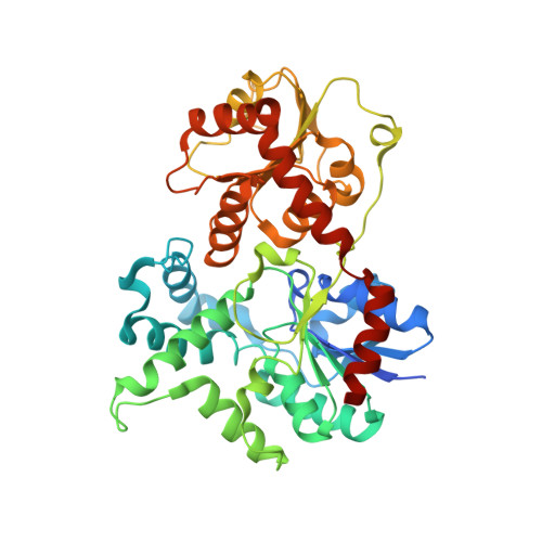
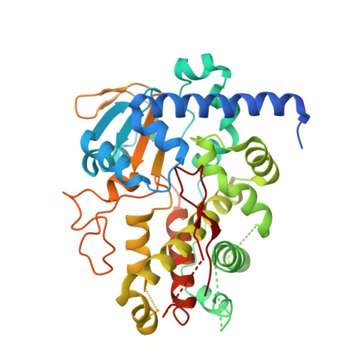
    In the biosynthesis of the clinically important antibiotic erythromycin D, the glycosyltransferase (GT) EryCIII, in concert with its partner EryCII, attaches a nucleotide-activated sugar to the macrolide scaffold with high specificity. To understand the role of EryCII, we have determined the crystal structure of the EryCIII·EryCII complex at 3.1 Å resolution. The structure reveals a heterotetramer with a distinctive, elongated quaternary organization. The EryCIII subunits form an extensive self-complementary dimer interface at the center of the complex, and the EryCII subunits lie on the periphery. EryCII binds in the vicinity of the putative macrolide binding site of EryCIII but does not make direct interactions with this site. Our biophysical and enzymatic data support a model in which EryCII stabilizes EryCIII and also functions as an allosteric activator of the GT.


  • Organizational Affiliation

    Department of Biochemistry, Cambridge University, Cambridge CB2 1GA, UK. mcm35@cam.ac.uk


Macromolecules
Find similar proteins by:  (by identity cutoff)  |  3D Structure
Entity ID: 1
MoleculeChains Sequence LengthOrganismDetailsImage
GLYCOSYLTRANSFERASE441Saccharopolyspora erythraea NRRL 2338Mutation(s): 0 
UniProt
Find proteins for A4F7P3 (Saccharopolyspora erythraea (strain ATCC 11635 / DSM 40517 / JCM 4748 / NBRC 13426 / NCIMB 8594 / NRRL 2338))
Explore A4F7P3 
Go to UniProtKB:  A4F7P3
Entity Groups  
Sequence Clusters30% Identity50% Identity70% Identity90% Identity95% Identity100% Identity
UniProt GroupA4F7P3
Sequence Annotations
Expand
  • Reference Sequence
Find similar proteins by:  (by identity cutoff)  |  3D Structure
Entity ID: 2
MoleculeChains Sequence LengthOrganismDetailsImage
DTDP-4-KETO-6-DEOXY-HEXOSE 3,4-ISOMERASE381Saccharopolyspora erythraea NRRL 2338Mutation(s): 0 
UniProt
Find proteins for A4F7P2 (Saccharopolyspora erythraea (strain ATCC 11635 / DSM 40517 / JCM 4748 / NBRC 13426 / NCIMB 8594 / NRRL 2338))
Explore A4F7P2 
Go to UniProtKB:  A4F7P2
Entity Groups  
Sequence Clusters30% Identity50% Identity70% Identity90% Identity95% Identity100% Identity
UniProt GroupA4F7P2
Sequence Annotations
Expand
  • Reference Sequence
Experimental Data & Validation

Experimental Data

  • Method: X-RAY DIFFRACTION
  • Resolution: 3.09 Å
  • R-Value Free: 0.262 
  • R-Value Work: 0.210 
  • R-Value Observed: 0.213 
  • Space Group: P 2 3
Unit Cell:
Length ( Å )Angle ( ˚ )
a = 141.891α = 90
b = 141.891β = 90
c = 141.891γ = 90
Software Package:
Software NamePurpose
PHENIXrefinement
XDSdata reduction
SCALAdata scaling
autoSHARPphasing

Structure Validation

View Full Validation Report


Entry History 

Deposition Data

Revision History  (Full details and data files)

  • Version 1.0: 2011-11-09
    Type: Initial release
  • Version 1.1: 2012-01-11
    Changes: Other
  • Version 1.2: 2015-04-01
    Changes: Database references