2YIE

Crystal structure of a F. nucleatum FMN riboswitch bound to FMN


Experimental Data Snapshot

  • Method: X-RAY DIFFRACTION
  • Resolution: 2.94 Å
  • R-Value Free: 0.231 
  • R-Value Work: 0.204 
  • R-Value Observed: 0.206 

wwPDB Validation   3D Report Full Report


Ligand Structure Quality Assessment 


This is version 1.2 of the entry. See complete history


Literature

Molecular Sensing by the Aptamer Domain of the Fmn Riboswitch: A General Model for Ligand Binding by Conformational Selection

Vicens, Q.Mondragon, E.Batey, R.T.

(2011) Nucleic Acids Res 39: 8586

  • DOI: https://doi.org/10.1093/nar/gkr565
  • Primary Citation of Related Structures:  
    2YIE, 2YIF

  • PubMed Abstract: 

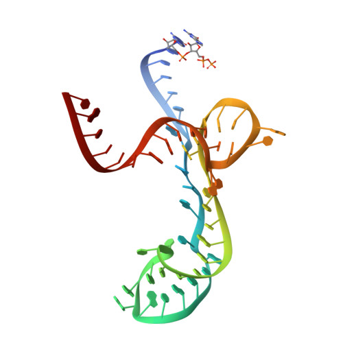
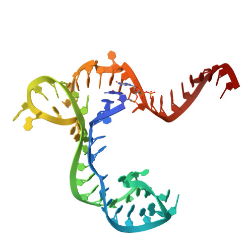
    Understanding the nature of the free state of riboswitch aptamers is important for illuminating common themes in gene regulation by riboswitches. Prior evidence indicated the flavin mononucleotide (FMN)-binding riboswitch aptamer adopted a 'bound-like' structure in absence of FMN, suggesting only local conformational changes upon ligand binding. In the scope of pinpointing the general nature of such changes at the nucleotide level, we performed SHAPE mapping experiments using the aptamer domain of two phylogenetic variants, both in absence and in presence of FMN. We also solved the crystal structures of one of these domains both free (3.3 Å resolution) and bound to FMN (2.95 Å resolution). Our comparative study reveals that structural rearrangements occurring upon binding are restricted to a few of the joining regions that form the binding pocket in both RNAs. This type of binding event with minimal structural perturbations is reminiscent of binding events by conformational selection encountered in other riboswitches and various RNAs.


  • Organizational Affiliation

    Department of Chemistry and Biochemistry, University of Colorado, Boulder, CO 80309-0215, USA.


Macromolecules
Find similar nucleic acids by:  (by identity cutoff)  |  3D Structure
Entity ID: 1
MoleculeChains LengthOrganismImage
FMN RIBOSWITCHA [auth X]54Fusobacterium nucleatum subsp. nucleatum ATCC 25586
Sequence Annotations
Expand
  • Reference Sequence
Find similar nucleic acids by:  (by identity cutoff)  |  3D Structure
Entity ID: 2
MoleculeChains LengthOrganismImage
FMN RIBOSWITCHB [auth Z]57Fusobacterium nucleatum subsp. nucleatum ATCC 25586
Sequence Annotations
Expand
  • Reference Sequence
Small Molecules
Ligands 3 Unique
IDChains Name / Formula / InChI Key2D Diagram3D Interactions
FMN
Query on FMN

Download Ideal Coordinates CCD File 
L [auth Z]FLAVIN MONONUCLEOTIDE
C17 H21 N4 O9 P
FVTCRASFADXXNN-SCRDCRAPSA-N
K
Query on K

Download Ideal Coordinates CCD File 
K [auth X],
S [auth Z]
POTASSIUM ION
K
NPYPAHLBTDXSSS-UHFFFAOYSA-N
MG
Query on MG

Download Ideal Coordinates CCD File 
C [auth X]
D [auth X]
E [auth X]
F [auth X]
G [auth X]
C [auth X],
D [auth X],
E [auth X],
F [auth X],
G [auth X],
H [auth X],
I [auth X],
J [auth X],
M [auth Z],
N [auth Z],
O [auth Z],
P [auth Z],
Q [auth Z],
R [auth Z]
MAGNESIUM ION
Mg
JLVVSXFLKOJNIY-UHFFFAOYSA-N
Experimental Data & Validation

Experimental Data

  • Method: X-RAY DIFFRACTION
  • Resolution: 2.94 Å
  • R-Value Free: 0.231 
  • R-Value Work: 0.204 
  • R-Value Observed: 0.206 
  • Space Group: P 31 2 1
Unit Cell:
Length ( Å )Angle ( ˚ )
a = 73.679α = 90
b = 73.679β = 90
c = 139.251γ = 120
Software Package:
Software NamePurpose
PHENIXrefinement
HKL-2000data scaling
CNSphasing

Structure Validation

View Full Validation Report


Ligand Structure Quality Assessment 


Entry History 

Deposition Data

Revision History  (Full details and data files)

  • Version 1.0: 2011-08-31
    Type: Initial release
  • Version 1.1: 2011-11-09
    Changes: Database references
  • Version 1.2: 2023-12-20
    Changes: Data collection, Database references, Derived calculations, Other, Refinement description