2YG2 | pdb_00002yg2

Structure of apolioprotein M in complex with Sphingosine 1-Phosphate


Experimental Data Snapshot

  • Method: X-RAY DIFFRACTION
  • Resolution: 1.70 Å
  • R-Value Free: 
    0.221 (Depositor), 0.230 (DCC) 
  • R-Value Work: 
    0.191 (Depositor), 0.205 (DCC) 
  • R-Value Observed: 
    0.192 (Depositor) 

Starting Model: experimental
View more details

wwPDB Validation 3D Report Full Report

Validation slider image for 2YG2

Ligand Structure Quality Assessment 


This is version 1.4 of the entry. See complete history

Literature

Endothelium-Protective Sphingosine-1-Phosphate Provided by Hdl-Associated Apolipoprotein M.

Christoffersen, C.Obinata, H.Kumaraswamy, S.B.Galvani, S.Ahnstrom, J.Sevvana, M.Egerer-Sieber, C.Muller, Y.A.Hla, T.Nielsen, L.B.Dahlback, B.

(2011) Proc Natl Acad Sci U S A 108: 9613

  • DOI: https://doi.org/10.1073/pnas.1103187108
  • Primary Citation Related Structures: 
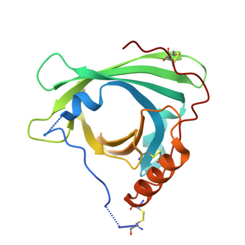
    2YG2

  • PubMed Abstract: 

    Protection of the endothelium is provided by circulating sphingosine-1-phosphate (S1P), which maintains vascular integrity. We show that HDL-associated S1P is bound specifically to both human and murine apolipoprotein M (apoM). Thus, isolated human ApoM(+) HDL contained S1P, whereas ApoM(-) HDL did not. Moreover, HDL in Apom(-/-) mice contains no S1P, whereas HDL in transgenic mice overexpressing human apoM has an increased S1P content. The 1.7-Å structure of the S1P-human apoM complex reveals that S1P interacts specifically with an amphiphilic pocket in the lipocalin fold of apoM. Human ApoM(+) HDL induced S1P(1) receptor internalization, downstream MAPK and Akt activation, endothelial cell migration, and formation of endothelial adherens junctions, whereas apoM(-) HDL did not. Importantly, lack of S1P in the HDL fraction of Apom(-/-) mice decreased basal endothelial barrier function in lung tissue. Our results demonstrate that apoM, by delivering S1P to the S1P(1) receptor on endothelial cells, is a vasculoprotective constituent of HDL.


  • Organizational Affiliation
    • Department of Clinical Biochemistry, Rigshospitalet, 2100 Copenhagen, Denmark.

Macromolecule Content 

  • Total Structure Weight: 39.43 kDa 
  • Atom Count: 2,763 
  • Modeled Residue Count: 314 
  • Deposited Residue Count: 344 
  • Unique protein chains: 1

Macromolecules

Find similar proteins by:|  3D Structure
Entity ID: 1
MoleculeChains  Sequence LengthOrganismDetailsImage
APOLIPOPROTEIN M
A, B
172Homo sapiensMutation(s): 0 
UniProt & NIH Common Fund Data Resources
Find proteins for O95445 (Homo sapiens)
Explore O95445 
Go to UniProtKB:  O95445
PHAROS:  O95445
GTEx:  ENSG00000204444 
Entity Groups
Sequence Clusters30% Identity50% Identity70% Identity90% Identity95% Identity100% Identity
UniProt GroupO95445
Sequence Annotations
Expand
Reference Sequence

Experimental Data & Validation

Experimental Data

  • Method: X-RAY DIFFRACTION
  • Resolution: 1.70 Å
  • R-Value Free:  0.221 (Depositor), 0.230 (DCC) 
  • R-Value Work:  0.191 (Depositor), 0.205 (DCC) 
  • R-Value Observed: 0.192 (Depositor) 
Space Group: P 31 2 1
Unit Cell:
Length ( Å )Angle ( ˚ )
a = 68.28α = 90
b = 68.28β = 90
c = 135.58γ = 120
Software Package:
Software NamePurpose
REFMACrefinement
XDSdata reduction
XSCALEdata scaling
PHASERphasing

Structure Validation

View Full Validation Report


Ligand Structure Quality Assessment 


Entry History 

Revision History  (Full details and data files)

  • Version 1.0: 2011-06-01
    Type: Initial release
  • Version 1.1: 2011-06-16
    Changes: Version format compliance
  • Version 1.2: 2011-07-13
    Changes: Version format compliance
  • Version 1.3: 2023-12-20
    Changes: Data collection, Database references, Derived calculations, Other, Refinement description
  • Version 1.4: 2024-11-13
    Changes: Structure summary