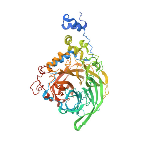
Crystal Structure of Inulosucrase from Lactobacillus: Insights Into the Substrate Specificity and Product Specificity of Gh68 Fructansucrases.
Pijning, T., Anwar, M.A., Boger, M., Dobruchowska, J.M., Leemhuis, H., Kralj, S., Dijkhuizen, L., Dijkstra, B.W.(2011) J Mol Biology 412: 80
- PubMed: 21801732
- DOI: https://doi.org/10.1016/j.jmb.2011.07.031
- Primary Citation of Related Structures:
2YFR, 2YFS, 2YFT - PubMed Abstract:
Fructansucrases (FSs) catalyze a transfructosylation reaction with sucrose as substrate to produce fructo-oligosaccharides and fructan polymers that contain either β-2,1 glycosidic linkages (inulin) or β-2,6 linkages (levan). Levan-synthesizing FSs (levansucrases) have been most extensively investigated, while detailed information on inulosucrases is limited. Importantly, the molecular basis of the different product specificities of levansucrases and inulosucrases is poorly understood. We have elucidated the three-dimensional structure of a truncated active bacterial GH68 inulosucrase, InuJ of Lactobacillus johnsonii NCC533 (residues 145-708), in its apo form, with a bound substrate (sucrose), and with a transfructosylation product. The sucrose binding pocket and the sucrose binding mode are virtually identical with those of GH68 levansucrases, confirming that both enzyme types use the same fully conserved structural framework for the binding and cleavage of the donor substrate sucrose in the active site. The binding mode of the first transfructosylation product 1-kestose (Fru-β(2-1)-Fru-α(2-1)-Glc, where Fru=fructose and Glc=glucose) in subsites -1 to +2 shows for the first time how inulin-type fructo-oligosaccharide bind in GH68 FS and how an inulin-type linkage can be formed. Surprisingly, observed interactions with the sugar in subsites +1 and +2 are provided by residues that are also present in levansucrases. The binding mode of 1-kestose and the presence of a more distant sucrose binding site suggest that residues beyond the +2 subsite, in particular residues from the nonconserved 1B-1C loop, determine product linkage type specificity in GH68 FSs.
- Laboratory of Biophysical Chemistry, Groningen Biomolecular Sciences and Biotechnology Institute, University of Groningen, Nijenborgh 7, 9747 AG Groningen, The Netherlands.
Organizational Affiliation:


