2YFH | pdb_00002yfh

Structure of a Chimeric Glutamate Dehydrogenase


Experimental Data Snapshot

  • Method: X-RAY DIFFRACTION
  • Resolution: 2.69 Å
  • R-Value Free: 
    0.282 (Depositor), 0.280 (DCC) 
  • R-Value Work: 
    0.215 (Depositor), 0.215 (DCC) 
  • R-Value Observed: 
    0.218 (Depositor) 

Starting Model: experimental
View more details

wwPDB Validation 3D Report Full Report

Validation slider image for 2YFH

This is version 1.4 of the entry. See complete history

Literature

Crystal Structure of a Chimaeric Bacterial Glutamate Dehydrogenase.

Oliveira, T.Sharkey, M.A.Engel, P.C.Khan, A.R.

(2016) Acta Crystallogr F Struct Biol Commun 72: 462

  • DOI: https://doi.org/10.1107/S2053230X16007305
  • Primary Citation Related Structures: 
    2YFH

  • PubMed Abstract: 

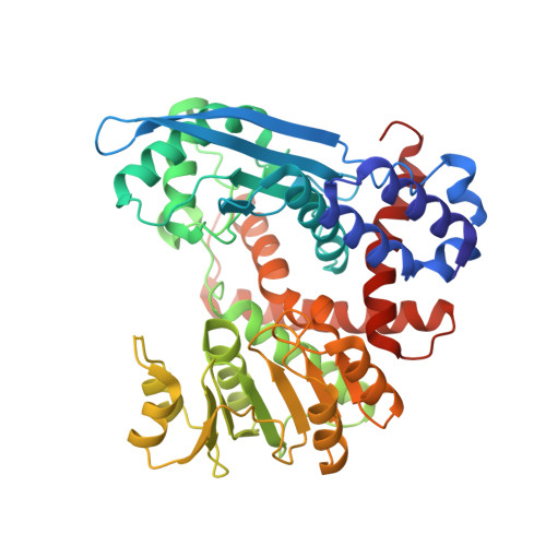
    Glutamate dehydrogenases (EC 1.4.1.2-4) catalyse the oxidative deamination of L-glutamate to α-ketoglutarate using NAD(P)(+) as a cofactor. The bacterial enzymes are hexameric, arranged with 32 symmetry, and each polypeptide consists of an N-terminal substrate-binding segment (domain I) followed by a C-terminal cofactor-binding segment (domain II). The catalytic reaction takes place in the cleft formed at the junction of the two domains. Distinct signature sequences in the nucleotide-binding domain have been linked to the binding of NAD(+) versus NADP(+), but they are not unambiguous predictors of cofactor preference. In the absence of substrate, the two domains move apart as rigid bodies, as shown by the apo structure of glutamate dehydrogenase from Clostridium symbiosum. Here, the crystal structure of a chimaeric clostridial/Escherichia coli enzyme has been determined in the apo state. The enzyme is fully functional and reveals possible determinants of interdomain flexibility at a hinge region following the pivot helix. The enzyme retains the preference for NADP(+) cofactor from the parent E. coli domain II, although there are subtle differences in catalytic activity.


  • Organizational Affiliation
    • School of Biochemistry and Immunology, Trinity College Dublin, Dublin 2, Ireland.

Macromolecule Content 

  • Total Structure Weight: 290.58 kDa 
  • Atom Count: 20,640 
  • Modeled Residue Count: 2,677 
  • Deposited Residue Count: 2,688 
  • Unique protein chains: 1

Macromolecules

Find similar proteins by:|  3D Structure
Entity ID: 1
MoleculeChains  Sequence LengthOrganismDetailsImage
GLUTAMATE DEHYDROGENASE, NAD-SPECIFIC GLUTAMATE DEHYDROGENASE, GLUTAMATE DEHYDROGENASE
A, B, C, D, E
A, B, C, D, E, F
448[Clostridium] symbiosumEscherichia coliMutation(s): 0 
EC: 1.4.1.4 (PDB Primary Data), 1.4.1.3 (PDB Primary Data), 1.4.1.2 (PDB Primary Data)
UniProt
Find proteins for P24295 (Clostridium symbiosum)
Explore P24295 
Go to UniProtKB:  P24295
Entity Groups
Sequence Clusters30% Identity50% Identity70% Identity90% Identity95% Identity100% Identity
UniProt GroupP24295
Sequence Annotations
Expand
Reference Sequence

Experimental Data & Validation

Experimental Data

  • Method: X-RAY DIFFRACTION
  • Resolution: 2.69 Å
  • R-Value Free:  0.282 (Depositor), 0.280 (DCC) 
  • R-Value Work:  0.215 (Depositor), 0.215 (DCC) 
  • R-Value Observed: 0.218 (Depositor) 
Space Group: P 1
Unit Cell:
Length ( Å )Angle ( ˚ )
a = 100.102α = 116.54
b = 113.859β = 101.54
c = 109.581γ = 104.09
Software Package:
Software NamePurpose
PHENIXrefinement
HKL-2000data reduction
SCALEPACKdata scaling
PHASERphasing

Structure Validation

View Full Validation Report


Entry History 

Deposition Data

Revision History  (Full details and data files)

  • Version 1.0: 2012-04-04
    Type: Initial release
  • Version 1.1: 2016-06-08
    Changes: Database references
  • Version 1.2: 2016-06-29
    Changes: Database references
  • Version 1.3: 2017-03-15
    Changes: Source and taxonomy
  • Version 1.4: 2023-12-20
    Changes: Data collection, Database references, Other, Refinement description