2YCY

TURKEY BETA1 ADRENERGIC RECEPTOR WITH STABILISING MUTATIONS AND BOUND ANTAGONIST CYANOPINDOLOL


Experimental Data Snapshot

  • Method: X-RAY DIFFRACTION
  • Resolution: 3.15 Å
  • R-Value Free: 0.292 
  • R-Value Work: 0.254 
  • R-Value Observed: 0.255 

wwPDB Validation   3D Report Full Report


Ligand Structure Quality Assessment 


This is version 1.3 of the entry. See complete history


Literature

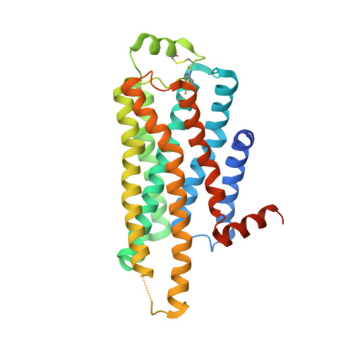
Two Distinct Conformations of Helix 6 Observed in Antagonist-Bound Structures of a {Beta}1- Adrenergic Receptor.

Moukhametzianov, R.Warne, T.Edwards, P.C.Serrano-Vega, M.J.Leslie, A.G.W.Tate, C.G.Schertler, G.F.X.

(2011) Proc Natl Acad Sci U S A 108: 8228

  • DOI: https://doi.org/10.1073/pnas.1100185108
  • Primary Citation of Related Structures:  
    2YCW, 2YCX, 2YCY, 2YCZ

  • PubMed Abstract: 
  • The β(1)-adrenergic receptor (β(1)AR) is a G-protein-coupled receptor whose inactive state structure was determined using a thermostabilized mutant (β(1)AR-M23). However, it was not thought to be in a fully inactivated state because there was no salt bridge between Arg139 and Glu285 linking the cytoplasmic ends of transmembrane helices 3 and 6 (the R(3 ...

    The β(1)-adrenergic receptor (β(1)AR) is a G-protein-coupled receptor whose inactive state structure was determined using a thermostabilized mutant (β(1)AR-M23). However, it was not thought to be in a fully inactivated state because there was no salt bridge between Arg139 and Glu285 linking the cytoplasmic ends of transmembrane helices 3 and 6 (the R(3.50) - D/E(6.30) "ionic lock"). Here we compare eight new structures of β(1)AR-M23, determined from crystallographically independent molecules in four different crystals with three different antagonists bound. These structures are all in the inactive R state and show clear electron density for cytoplasmic loop 3 linking transmembrane helices 5 and 6 that had not been seen previously. Despite significantly different crystal packing interactions, there are only two distinct conformations of the cytoplasmic end of helix 6, bent and straight. In the bent conformation, the Arg139-Glu285 salt bridge is present, as in the crystal structure of dark-state rhodopsin. The straight conformation, observed in previously solved structures of β-receptors, results in the ends of helices 3 and 6 being too far apart for the ionic lock to form. In the bent conformation, the R(3.50)-E(6.30) distance is significantly longer than in rhodopsin, suggesting that the interaction is also weaker, which could explain the high basal activity in β(1)AR compared to rhodopsin. Many mutations that increase the constitutive activity of G-protein-coupled receptors are found in the bent region at the cytoplasmic end of helix 6, supporting the idea that this region plays an important role in receptor activation.


    Organizational Affiliation

    Medical Research Council Laboratory of Molecular Biology, Hills Road, Cambridge CB2 0QH, United Kingdom.


Macromolecules
Find similar proteins by:  (by identity cutoff)  |  3D Structure
Entity ID: 1
MoleculeChainsSequence LengthOrganismDetailsImage
BETA-1 ADRENERGIC RECEPTORA, B313Meleagris gallopavoMutation(s): 8 
Gene Names: ADRB1
Membrane Entity: Yes 
UniProt
Find proteins for P07700 (Meleagris gallopavo)
Explore P07700 
Go to UniProtKB:  P07700
Entity Groups  
Sequence Clusters30% Identity50% Identity70% Identity90% Identity95% Identity100% Identity
UniProt GroupP07700
Protein Feature View
Expand
  • Reference Sequence
Small Molecules
Ligands 3 Unique
IDChainsName / Formula / InChI Key2D Diagram3D Interactions
SOG
Query on SOG

Download Ideal Coordinates CCD File 
D [auth A],
E [auth A],
G [auth B],
H [auth B],
I [auth B]
octyl 1-thio-beta-D-glucopyranoside
C14 H28 O5 S
CGVLVOOFCGWBCS-RGDJUOJXSA-N
 Ligand Interaction
P32
Query on P32

Download Ideal Coordinates CCD File 
C [auth A],
F [auth B]
4-{[(2S)-3-(tert-butylamino)-2-hydroxypropyl]oxy}-3H-indole-2-carbonitrile
C16 H21 N3 O2
CQEFAUFOQSCRMZ-LBPRGKRZSA-N
 Ligand Interaction
NA
Query on NA

Download Ideal Coordinates CCD File 
J [auth B]SODIUM ION
Na
FKNQFGJONOIPTF-UHFFFAOYSA-N
 Ligand Interaction
Experimental Data & Validation

Experimental Data

  • Method: X-RAY DIFFRACTION
  • Resolution: 3.15 Å
  • R-Value Free: 0.292 
  • R-Value Work: 0.254 
  • R-Value Observed: 0.255 
  • Space Group: C 1 2 1
Unit Cell:
Length ( Å )Angle ( ˚ )
a = 184.76α = 90
b = 56.161β = 111.27
c = 89.778γ = 90
Software Package:
Software NamePurpose
REFMACrefinement
MOSFLMdata reduction
SCALAdata scaling
PHASERphasing

Structure Validation

View Full Validation Report


Ligand Structure Quality Assessment 


Entry History 

Deposition Data

Revision History  (Full details and data files)

  • Version 1.0: 2011-06-08
    Type: Initial release
  • Version 1.1: 2011-07-13
    Changes: Version format compliance
  • Version 1.2: 2019-04-03
    Changes: Data collection, Other, Source and taxonomy
  • Version 1.3: 2020-07-29
    Type: Remediation
    Reason: Carbohydrate remediation
    Changes: Derived calculations, Other, Structure summary