2Y9H | pdb_00002y9h

Structure A of CRISPR endoribonuclease Cse3 bound to 19 nt RNA


Experimental Data Snapshot

  • Method: X-RAY DIFFRACTION
  • Resolution: 2.50 Å
  • R-Value Free: 
    0.289 (Depositor), 0.292 (DCC) 
  • R-Value Work: 
    0.231 (Depositor), 0.235 (DCC) 
  • R-Value Observed: 
    0.232 (Depositor) 

Starting Model: experimental
View more details

wwPDB Validation 3D Report Full Report

Validation slider image for 2Y9H

This is version 1.4 of the entry. See complete history

Literature

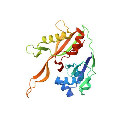
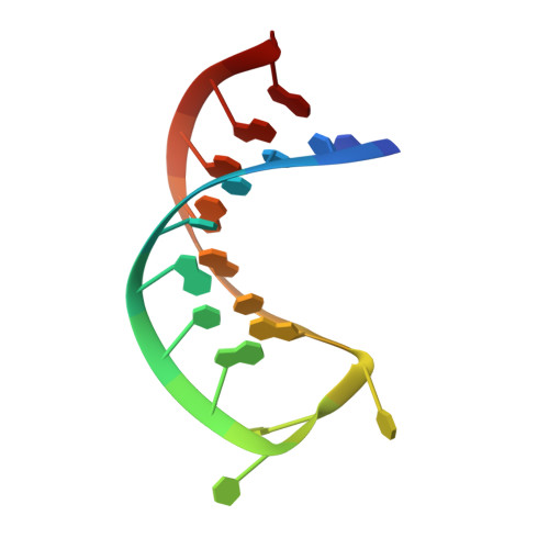
An RNA-Induced Conformational Change Required for Crispr RNA Cleavage by the Endoribonuclease Cse3.

Sashital, D.G.Jinek, M.Doudna, J.A.

(2011) Nat Struct Mol Biol 18: 680

  • DOI: https://doi.org/10.1038/nsmb.2043
  • Primary Citation Related Structures: 
    2Y8W, 2Y8Y, 2Y9H

  • PubMed Abstract: 

    Clustered regularly interspaced short palindromic repeat (CRISPR) chromosomal loci found in prokaryotes provide an adaptive immune system against bacteriophages and plasmids. CRISPR-specific endoRNases produce short RNA molecules (crRNAs) from CRISPR transcripts, which harbor sequences complementary to invasive nucleic acid elements and ensure their selective targeting by CRISPR-associated (Cas) proteins. The extreme sequence divergence of CRISPR-specific endoRNases and their RNA substrates has obscured homology-based comparison of RNA recognition and cleavage mechanisms. Here, we show that Cse3 type CRISPR-specific endoRNases bind a hairpin structure and residues downstream of the cleavage site within the repetitive segment of cognate CRISPR RNA. Cocrystal structures of Cse3-RNA complexes reveal an RNA-induced conformational change in the enzyme active site that aligns the RNA strand for site-specific cleavage. These studies provide insight into a catalytically essential RNA recognition mechanism by a large class of CRISPR-related endoRNases.


  • Organizational Affiliation
    • Department of Molecular and Cell Biology, University of California, Berkeley, California, USA.

Macromolecule Content 

  • Total Structure Weight: 240.96 kDa 
  • Atom Count: 15,663 
  • Modeled Residue Count: 1,740 
  • Deposited Residue Count: 1,872 
  • Unique protein chains: 1
  • Unique nucleic acid chains: 1

Macromolecules


Find similar proteins by:|  3D Structure
Entity ID: 1
MoleculeChains  Sequence LengthOrganismDetailsImage
CSE3
A, C, E, G, I
A, C, E, G, I, K, M, O
215Thermus thermophilus HB8Mutation(s): 0 
EC: 3.1
UniProt
Find proteins for Q53WG9 (Thermus thermophilus (strain ATCC 27634 / DSM 579 / HB8))
Explore Q53WG9 
Go to UniProtKB:  Q53WG9
Entity Groups
Sequence Clusters30% Identity50% Identity70% Identity90% Identity95% Identity100% Identity
UniProt GroupQ53WG9
Sequence Annotations
Expand
Reference Sequence
Find similar nucleic acids by:  Sequence
Entity ID: 2
MoleculeChains LengthOrganismImage
5'-R(*UP*CP*CP*CP*CP*AP*CP*GP*CP*GP*UP*GP*UP*GP *GP*GP*DGP*AP*UP)-3'
B, D, F, H, J
B, D, F, H, J, L, N, P
19N/A
Sequence Annotations
Expand
Reference Sequence

Experimental Data & Validation

Experimental Data

  • Method: X-RAY DIFFRACTION
  • Resolution: 2.50 Å
  • R-Value Free:  0.289 (Depositor), 0.292 (DCC) 
  • R-Value Work:  0.231 (Depositor), 0.235 (DCC) 
  • R-Value Observed: 0.232 (Depositor) 
Space Group: P 1 21 1
Unit Cell:
Length ( Å )Angle ( ˚ )
a = 85.86α = 90
b = 149.81β = 94.6
c = 87.26γ = 90
Software Package:
Software NamePurpose
PHENIXrefinement
XDSdata reduction
XSCALEdata scaling
PHASERphasing

Structure Validation

View Full Validation Report


Entry History 

Deposition Data

Revision History  (Full details and data files)

  • Version 1.0: 2011-05-18
    Type: Initial release
  • Version 1.1: 2011-06-09
    Changes: Version format compliance
  • Version 1.2: 2011-07-13
    Changes: Version format compliance
  • Version 1.3: 2022-11-02
    Changes: Database references, Other
  • Version 1.4: 2024-01-31
    Changes: Data collection, Refinement description