Insights Into the Function of the Crm1 Cofactor Ranbp3 from the Structure of its Ran-Binding Domain
Langer, K., Dian, C., Rybin, V., Muller, C.W., Petosa, C.(2011) PLoS One 6: 17011
- PubMed: 21364925 Search on PubMedSearch on PubMed Central
- DOI: https://doi.org/10.1371/journal.pone.0017011
- Primary Citation Related Structures:
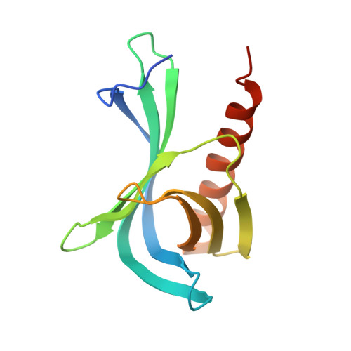
2Y8F, 2Y8G - PubMed Abstract:
Proteins bearing a leucine-rich nuclear export signal (NES) are exported from the nucleus by the transport factor CRM1, which forms a cooperative ternary complex with the NES-bearing cargo and with the small GTPase Ran. CRM1-mediated export is regulated by RanBP3, a Ran-interacting nuclear protein. Unlike the related proteins RanBP1 and RanBP2, which promote disassembly of the export complex in the cytosol, RanBP3 acts as a CRM1 cofactor, enhancing NES export by stabilizing the export complex in the nucleus. RanBP3 also alters the cargo selectivity of CRM1, promoting recognition of the NES of HIV-1 Rev and of other cargos while deterring recognition of the import adaptor protein Snurportin1. Here we report the crystal structure of the Ran-binding domain (RBD) from RanBP3 and compare it to RBD structures from RanBP1 and RanBP2 in complex with Ran and CRM1. Differences among these structures suggest why RanBP3 binds Ran with unusually low affinity, how RanBP3 modulates the cargo selectivity of CRM1, and why RanBP3 promotes assembly rather than disassembly of the export complex. The comparison of RBD structures thus provides an insight into the functional diversity of Ran-binding proteins.
- Structural and Computational Biology Unit, European Molecular Biology Laboratory, Heidelberg, Germany.
Organizational Affiliation: