2Y7L | pdb_00002y7l

Structure of N-terminal domain of Candida albicans Als9-2 in complex with human fibrinogen gamma peptide


Experimental Data Snapshot

  • Method: X-RAY DIFFRACTION
  • Resolution: 1.49 Å
  • R-Value Free: 
    0.224 (Depositor), 0.224 (DCC) 
  • R-Value Work: 
    0.196 (Depositor), 0.196 (DCC) 
  • R-Value Observed: 
    0.198 (Depositor) 

Starting Model: experimental
View more details

wwPDB Validation 3D Report Full Report

Validation slider image for 2Y7L

This is version 1.2 of the entry. See complete history

Literature

Structural Basis for the Broad Specificity to Host- Cell Ligands by the Pathogenic Fungus Candida Albicans.

Salgado, P.S.Yan, R.Taylor, J.D.Burchell, L.Jones, R.Hoyer, L.L.Matthews, S.J.Simpson, P.J.Cota, E.

(2011) Proc Natl Acad Sci U S A 108: 15775

  • DOI: https://doi.org/10.1073/pnas.1103496108
  • Primary Citation Related Structures: 
    2Y7L, 2Y7M, 2Y7N, 2Y7O, 2YLH

  • PubMed Abstract: 

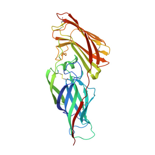
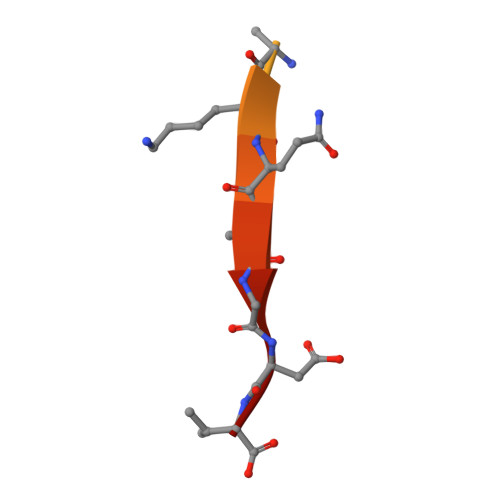
    Candida albicans is the most prevalent fungal pathogen in humans and a major source of life-threatening nosocomial infections. The Als (agglutinin-like sequence) glycoproteins are an important virulence factor for this fungus and have been associated with binding of host-cell surface proteins and small peptides of random sequence, the formation of biofilms and amyloid fibers. High-resolution structures of N-terminal Als adhesins (NT-Als; up to 314 amino acids) show that ligand recognition relies on a motif capable of binding flexible C termini of peptides in extended conformation. Central to this mechanism is an invariant lysine that recognizes the C-terminal carboxylate of ligands at the end of a deep-binding cavity. In addition to several protein-peptide interactions, a network of water molecules runs parallel to one side of the ligand and contributes to the recognition of diverse peptide sequences. These data establish NT-Als adhesins as a separate family of peptide-binding proteins and an unexpected adhesion system for primary, widespread protein-protein interactions at the Candida/host-cell interface.


  • Organizational Affiliation
    • Division of Molecular Biosciences, Imperial College London, Exhibition Road, South Kensington SW7 2AZ, United Kingdom.

Macromolecule Content 

  • Total Structure Weight: 35.29 kDa 
  • Atom Count: 2,739 
  • Modeled Residue Count: 319 
  • Deposited Residue Count: 329 
  • Unique protein chains: 2

Macromolecules

Find similar proteins by:|  3D Structure
Entity ID: 1
MoleculeChains  Sequence LengthOrganismDetailsImage
AGGLUTININ-LIKE ALS9 PROTEIN312Candida albicansMutation(s): 0 
UniProt
Find proteins for A0A1D8PQ86 (Candida albicans (strain SC5314 / ATCC MYA-2876))
Explore A0A1D8PQ86 
Go to UniProtKB:  A0A1D8PQ86
Entity Groups
Sequence Clusters30% Identity50% Identity70% Identity90% Identity95% Identity100% Identity
UniProt GroupA0A1D8PQ86
Sequence Annotations
Expand
Reference Sequence
Find similar proteins by:  Sequence   |   3D Structure  
Entity ID: 2
MoleculeChains  Sequence LengthOrganismDetailsImage
FIBRINOGEN GAMMA CHAIN, ISOFORM CRA_A17Homo sapiensMutation(s): 0 
UniProt & NIH Common Fund Data Resources
Find proteins for P02679 (Homo sapiens)
Explore P02679 
Go to UniProtKB:  P02679
PHAROS:  P02679
GTEx:  ENSG00000171557 
Entity Groups
Sequence Clusters30% Identity50% Identity70% Identity90% Identity95% Identity100% Identity
UniProt GroupP02679
Sequence Annotations
Expand
Reference Sequence

Experimental Data & Validation

Experimental Data

  • Method: X-RAY DIFFRACTION
  • Resolution: 1.49 Å
  • R-Value Free:  0.224 (Depositor), 0.224 (DCC) 
  • R-Value Work:  0.196 (Depositor), 0.196 (DCC) 
  • R-Value Observed: 0.198 (Depositor) 
Space Group: P 21 21 21
Unit Cell:
Length ( Å )Angle ( ˚ )
a = 35.581α = 90
b = 69.273β = 90
c = 122.223γ = 90
Software Package:
Software NamePurpose
REFMACrefinement
DENZOdata reduction
SCALEPACKdata scaling
PHASERphasing

Structure Validation

View Full Validation Report


Entry History 

Deposition Data

Revision History  (Full details and data files)

  • Version 1.0: 2011-10-05
    Type: Initial release
  • Version 1.1: 2023-12-20
    Changes: Data collection, Database references, Other, Refinement description
  • Version 1.2: 2024-11-06
    Changes: Structure summary