2Y6M

Crystal structure of EphA4 kinase domain


Experimental Data Snapshot

  • Method: X-RAY DIFFRACTION
  • Resolution: 1.70 Å
  • R-Value Free: 0.203 
  • R-Value Work: 0.175 
  • R-Value Observed: 0.176 

wwPDB Validation   3D Report Full Report


This is version 1.3 of the entry. See complete history


Literature

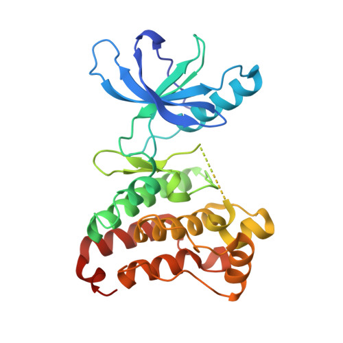
Crystal Structure of the Epha4 Protein Tyrosine Kinase Domain in the Apo- and Dasatinib-Bound State.

Farenc, C.J.A.Celie, P.H.N.Tensen, C.P.Siegal, G.

(2011) FEBS Lett 585: 3593

  • DOI: https://doi.org/10.1016/j.febslet.2011.10.028
  • Primary Citation of Related Structures:  
    2Y6M, 2Y6O

  • PubMed Abstract: 

    The Eph family of receptor tyrosine kinases regulates diverse cellular processes while the over-expression of a member of this family, EphA4, has been reported in a variety of malignant carcinomas. To gain insight into molecular mechanisms and to facilitate structure-based inhibitor design, we solved the crystal structure of the native EphA4 kinase domain in both the apo and dasatinib bound forms. Analysis of the two structures provides insight into structural features of inhibitor binding and revealed a hydrophobic back-pocket in the ATP- binding site of EphA4 which was previously unidentified. The structures suggest a route towards development of novel and specific inhibitors.


  • Organizational Affiliation

    Protein Chemistry Group, Leiden Institute of Chemistry, Leiden University, Leiden, The Netherlands.


Macromolecules
Find similar proteins by:  (by identity cutoff)  |  3D Structure
Entity ID: 1
MoleculeChains Sequence LengthOrganismDetailsImage
EPHRIN TYPE-A RECEPTOR 4291Mus musculusMutation(s): 0 
EC: 2.7.10.1
UniProt
Find proteins for Q03137 (Mus musculus)
Explore Q03137 
Go to UniProtKB:  Q03137
Entity Groups  
Sequence Clusters30% Identity50% Identity70% Identity90% Identity95% Identity100% Identity
UniProt GroupQ03137
Sequence Annotations
Expand
  • Reference Sequence
Experimental Data & Validation

Experimental Data

  • Method: X-RAY DIFFRACTION
  • Resolution: 1.70 Å
  • R-Value Free: 0.203 
  • R-Value Work: 0.175 
  • R-Value Observed: 0.176 
  • Space Group: P 21 21 21
Unit Cell:
Length ( Å )Angle ( ˚ )
a = 32.513α = 90
b = 91.045β = 90
c = 97.738γ = 90
Software Package:
Software NamePurpose
PHENIXrefinement
XDSdata reduction
XDSdata scaling
AMoREphasing

Structure Validation

View Full Validation Report


Entry History 

Deposition Data

Revision History  (Full details and data files)

  • Version 1.0: 2011-11-02
    Type: Initial release
  • Version 1.1: 2011-11-09
    Changes: Database references
  • Version 1.2: 2011-11-23
    Changes: Database references
  • Version 1.3: 2023-12-20
    Changes: Data collection, Database references, Derived calculations, Other, Refinement description