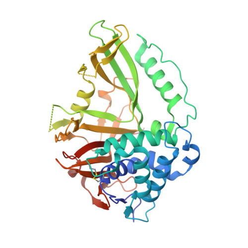
2Y6E

Structure of the D1D2 domain of USP4, the conserved catalytic domain


Experimental Data Snapshot

  • Method: X-RAY DIFFRACTION
  • Resolution: 2.40 Å
  • R-Value Free: 0.210 
  • R-Value Work: 0.178 
  • R-Value Observed: 0.180 

Starting Model: experimental
View more details

wwPDB Validation   3D Report Full Report


This is version 1.5 of the entry. See complete history


Literature

The Dusp-Ubl Domain of Usp4 Enhances its Catalytic Efficiency by Promoting Ubiquitin Exchange.

Clerici, M.Luna-Vargas, M.P.A.Faesen, A.C.Sixma, T.K.

(2014) Nat Commun 5: 5399

  • DOI: https://doi.org/10.1038/ncomms6399
  • Primary Citation of Related Structures:  
    2Y6E

  • PubMed Abstract: 

    Ubiquitin-specific protease USP4 is emerging as an important regulator of cellular pathways, including the TGF-β response, NF-κB signalling and splicing, with possible roles in cancer. Here we show that USP4 has its catalytic triad arranged in a productive conformation. Nevertheless, it requires its N-terminal DUSP-Ubl domain to achieve full catalytic turnover. Pre-steady-state kinetics measurements reveal that USP4 catalytic domain activity is strongly inhibited by slow dissociation of ubiquitin after substrate hydrolysis. The DUSP-Ubl domain is able to enhance ubiquitin dissociation, hence promoting efficient turnover. In a mechanism that requires all USP4 domains, binding of the DUSP-Ubl domain promotes a change of a switching loop near the active site. This 'allosteric regulation of product discharge' provides a novel way of regulating deubiquitinating enzymes that may have relevance for other enzyme classes.


  • Organizational Affiliation

    Department of Biochemistry, The Netherlands Cancer Institute, Plesmanlaan 121, 1066CX Amsterdam, The Netherlands.


Macromolecules
Find similar proteins by:  (by identity cutoff)  |  3D Structure
Entity ID: 1
MoleculeChains Sequence LengthOrganismDetailsImage
UBIQUITIN CARBOXYL-TERMINAL HYDROLASE 4
A, B, C, D, E
A, B, C, D, E, F
367Homo sapiensMutation(s): 0 
EC: 3.1.2.15 (PDB Primary Data), 3.4.19.12 (UniProt)
UniProt & NIH Common Fund Data Resources
Find proteins for Q13107 (Homo sapiens)
Explore Q13107 
Go to UniProtKB:  Q13107
PHAROS:  Q13107
GTEx:  ENSG00000114316 
Entity Groups  
Sequence Clusters30% Identity50% Identity70% Identity90% Identity95% Identity100% Identity
UniProt GroupQ13107
Sequence Annotations
Expand
  • Reference Sequence
Small Molecules
Ligands 2 Unique
IDChains Name / Formula / InChI Key2D Diagram3D Interactions
SO4
Query on SO4

Download Ideal Coordinates CCD File 
H [auth A],
K [auth C]
SULFATE ION
O4 S
QAOWNCQODCNURD-UHFFFAOYSA-L
ZN
Query on ZN

Download Ideal Coordinates CCD File 
G [auth A]
I [auth B]
J [auth C]
L [auth D]
M [auth E]
G [auth A],
I [auth B],
J [auth C],
L [auth D],
M [auth E],
N [auth F]
ZINC ION
Zn
PTFCDOFLOPIGGS-UHFFFAOYSA-N
Modified Residues  1 Unique
IDChains TypeFormula2D DiagramParent
CME
Query on CME
A, B, C, D, E
A, B, C, D, E, F
L-PEPTIDE LINKINGC5 H11 N O3 S2CYS
Experimental Data & Validation

Experimental Data

  • Method: X-RAY DIFFRACTION
  • Resolution: 2.40 Å
  • R-Value Free: 0.210 
  • R-Value Work: 0.178 
  • R-Value Observed: 0.180 
  • Space Group: P 21 21 21
Unit Cell:
Length ( Å )Angle ( ˚ )
a = 110.5α = 90
b = 151.03β = 90
c = 178.67γ = 90
Software Package:
Software NamePurpose
BUSTERrefinement
MOSFLMdata reduction
SCALAdata scaling
PHASERphasing

Structure Validation

View Full Validation Report


Entry History 

Deposition Data

Revision History  (Full details and data files)

  • Version 1.0: 2011-04-06
    Type: Initial release
  • Version 1.1: 2014-01-22
    Changes: Database references, Refinement description, Version format compliance
  • Version 1.2: 2014-10-22
    Changes: Database references
  • Version 1.3: 2014-12-03
    Changes: Database references, Structure summary
  • Version 1.4: 2023-12-20
    Changes: Data collection, Database references, Derived calculations, Other, Refinement description
  • Version 1.5: 2024-11-06
    Changes: Structure summary