2Y5M

STRUCTURE OF LINEAR GRAMICIDIN D OBTAINED USING TYPE I CRYSTALS GROWN IN A 7.7 MONOACYLGLYCEROL LIPID CUBIC PHASE.


Experimental Data Snapshot

  • Method: X-RAY DIFFRACTION
  • Resolution: 1.08 Å
  • R-Value Free: 0.155 
  • R-Value Work: 0.130 
  • R-Value Observed: 0.132 

wwPDB Validation   3D Report Full Report


This is version 2.1 of the entry. See complete history


Literature

Membrane Protein Crystallization in Lipidic Mesophases. Hosting Lipid Effects on the Crystallization and Structure of a Transmembrane Peptide

Hoefer, N.Aragao, D.Lyons, J.Caffrey, M.

(2011) Cryst Growth Des 11: 1182

  • DOI: https://doi.org/10.1021/cg101384p
  • Primary Citation of Related Structures:  
    2Y5M, 2Y6N

  • PubMed Abstract: 

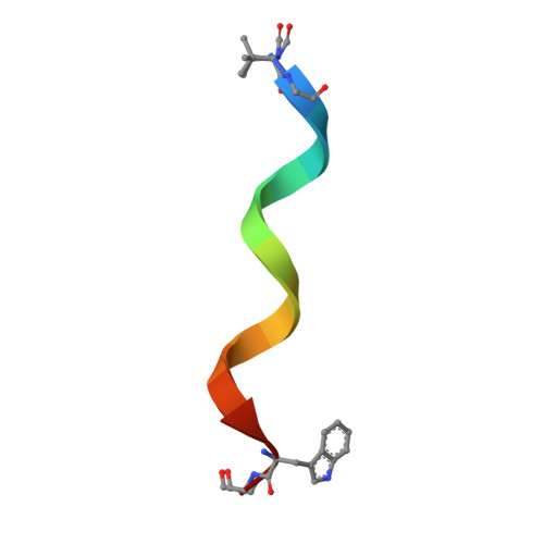
    Gramicidin is an apolar pentadecapeptide antibiotic consisting of alternating D-and L-amino acids. It functions, in part, by creating pores in membranes of susceptible cells rendering them leaky to monovalent cations. The peptide should be able to traverse the host membrane either as a double stranded, intertwined double helix (DSDH) or as a head-to-head single stranded helix (HHSH). Current structure models are based on macromolecular X-ray crystallography (MX) and nuclear magnetic resonance (NMR). However, the HHSH form has only been observed by NMR. The shape and size of the different gramicidin conformations differ. We speculated therefore that reconstituting it into a lipidic mesophase with bilayers of different microstructures would preferentially stabilize one form over the other. By using such mesophases for in meso crystallogenesis the expectation was that at least one would generate crystals of gramicidin in the HHSH form for structure determination by MX. This was tested using commercial and in-house synthesised lipids that support in meso crystallogenesis. Lipid acyl chain lengths were varied from 14 to 18 carbons to provide mesophases with a range of bilayer thicknesses. Unexpectedly, all lipids produced high quality, structure-grade crystals with gramicidin only in the DSDH conformation.


  • Organizational Affiliation

    Membrane Structural and Functional Biology Group, School of Biochemistry and Immunology, and School of Medicine, Trinity College, Dublin.


Macromolecules

Find similar proteins by:  Sequence   |   3D Structure  

Entity ID: 1
MoleculeChains Sequence LengthOrganismDetailsImage
VAL-GRAMICIDIN A
A, B, C, D, E
A, B, C, D, E, F
16Brevibacillus brevisMutation(s): 0 
Entity Groups  
Sequence Clusters30% Identity50% Identity70% Identity90% Identity95% Identity100% Identity
Sequence Annotations
Expand
  • Reference Sequence
Small Molecules
Modified Residues  1 Unique
IDChains TypeFormula2D DiagramParent
FVA
Query on FVA
A, B, C, D, E
A, B, C, D, E, F
L-PEPTIDE LINKINGC6 H11 N O3VAL
Experimental Data & Validation

Experimental Data

  • Method: X-RAY DIFFRACTION
  • Resolution: 1.08 Å
  • R-Value Free: 0.155 
  • R-Value Work: 0.130 
  • R-Value Observed: 0.132 
  • Space Group: P 1 21 1
Unit Cell:
Length ( Å )Angle ( ˚ )
a = 30.639α = 90
b = 62.794β = 100.02
c = 30.664γ = 90
Software Package:
Software NamePurpose
REFMACrefinement
HKL-2000data reduction
HKL-2000data scaling
PHASERphasing

Structure Validation

View Full Validation Report


Entry History 

Deposition Data

Revision History  (Full details and data files)

  • Version 1.0: 2011-05-11
    Type: Initial release
  • Version 1.1: 2013-01-30
    Changes: Database references, Version format compliance
  • Version 1.2: 2019-03-06
    Changes: Data collection, Derived calculations, Experimental preparation, Other
  • Version 1.3: 2023-03-29
    Changes: Database references, Derived calculations, Other, Structure summary
  • Version 2.0: 2023-11-15
    Changes: Atomic model, Data collection, Derived calculations
  • Version 2.1: 2024-02-07
    Changes: Refinement description