2Y4Z | pdb_00002y4z

Structure of the amino-terminal capsid restriction escape mutation N- MLV L10W


Experimental Data Snapshot

  • Method: X-RAY DIFFRACTION
  • Resolution: 2.00 Å
  • R-Value Free: 
    0.244 (Depositor), 0.238 (DCC) 
  • R-Value Work: 
    0.174 (Depositor), 0.170 (DCC) 
  • R-Value Observed: 
    0.177 (Depositor) 

Starting Model: experimental
View more details

wwPDB Validation 3D Report Full Report

Validation slider image for 2Y4Z

This is version 1.1 of the entry. See complete history

Literature

Novel Escape Mutants Suggest an Extensive Trim5Alpha Binding Site Spanning the Entire Outer Surface of the Murine Leukemia Virus Capsid Protein.

Ohkura, S.Goldstone, D.C.Yap, M.W.Holden-Dye, K.Taylor, I.A.Stoye, J.P.

(2011) PLoS Pathog 7: 2011

  • DOI: https://doi.org/10.1371/journal.ppat.1002011
  • Primary Citation Related Structures: 
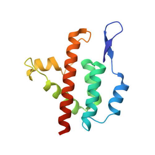
    2Y4Z

  • PubMed Abstract: 

    After entry into target cells, retroviruses encounter the host restriction factors such as Fv1 and TRIM5α. While it is clear that these factors target retrovirus capsid proteins (CA), recognition remains poorly defined in the absence of structural information. To better understand the binding interaction between TRIM5α and CA, we selected a panel of novel N-tropic murine leukaemia virus (N-MLV) escape mutants by a serial passage of replication competent N-MLV in rhesus macaque TRIM5α (rhTRIM5α)-positive cells using a small percentage of unrestricted cells to allow multiple rounds of virus replication. The newly identified mutations, many of which involve changes in charge, are distributed over the outer 'top' surface of N-MLV CA, including the N-terminal β-hairpin, and map up to 29 A(o) apart. Biological characterisation with a number of restriction factors revealed that only one of the new mutations affects restriction by human TRIM5α, indicating significant differences in the binding interaction between N-MLV and the two TRIM5αs, whereas three of the mutations result in dual sensitivity to Fv1(n) and Fv1(b). Structural studies of two mutants show that no major changes in the overall CA conformation are associated with escape from restriction. We conclude that interactions involving much, if not all, of the surface of CA are vital for TRIM5α binding.


  • Organizational Affiliation
    • Division of Virology, MRC National Institute for Medical Research, London, United Kingdom.

Macromolecule Content 

  • Total Structure Weight: 16.2 kDa 
  • Atom Count: 1,193 
  • Modeled Residue Count: 135 
  • Deposited Residue Count: 140 
  • Unique protein chains: 1

Macromolecules

Find similar proteins by:|  3D Structure
Entity ID: 1
MoleculeChains  Sequence LengthOrganismDetailsImage
CAPSID PROTEIN P30140Murine leukemia virusMutation(s): 1 
UniProt
Find proteins for P03336 (AKV murine leukemia virus)
Explore P03336 
Go to UniProtKB:  P03336
Entity Groups
Sequence Clusters30% Identity50% Identity70% Identity90% Identity95% Identity100% Identity
UniProt GroupP03336
Sequence Annotations
Expand
Reference Sequence

Small Molecules

Ligands 1 Unique
IDChains Name / Formula / InChI Key2D Diagram3D Interactions
GOL

Query on GOL


Download:Ideal Coordinates CCD File
B [auth A]GLYCEROL
C3 H8 O3
PEDCQBHIVMGVHV-UHFFFAOYSA-N

Experimental Data & Validation

Experimental Data

  • Method: X-RAY DIFFRACTION
  • Resolution: 2.00 Å
  • R-Value Free:  0.244 (Depositor), 0.238 (DCC) 
  • R-Value Work:  0.174 (Depositor), 0.170 (DCC) 
  • R-Value Observed: 0.177 (Depositor) 
Space Group: C 1 2 1
Unit Cell:
Length ( Å )Angle ( ˚ )
a = 71.88α = 90
b = 33.647β = 111.91
c = 59.32γ = 90
Software Package:
Software NamePurpose
PHENIXrefinement
DENZOdata reduction
SCALEPACKdata scaling
PHASERphasing

Structure Validation

View Full Validation Report


Entry History 

Deposition Data

Revision History  (Full details and data files)

  • Version 1.0: 2011-11-23
    Type: Initial release
  • Version 1.1: 2023-12-20
    Changes: Data collection, Database references, Derived calculations, Other, Refinement description