2Y3R | pdb_00002y3r

Structure of the tirandamycin-bound FAD-dependent tirandamycin oxidase TamL in P21 space group


Experimental Data Snapshot

  • Method: X-RAY DIFFRACTION
  • Resolution: 1.79 Å
  • R-Value Free: 
    0.219 (Depositor), 0.218 (DCC) 
  • R-Value Work: 
    0.146 (Depositor), 0.146 (DCC) 
  • R-Value Observed: 
    0.150 (Depositor) 

Starting Model: experimental
View more details

wwPDB Validation 3D Report Full Report

Validation slider image for 2Y3R

Ligand Structure Quality Assessment 


This is version 1.4 of the entry. See complete history

Literature

Tirandamycin Biosynthesis is Mediated by Co-Dependent Oxidative Enzymes

Carlson, J.C.Li, S.Gunatilleke, S.S.Anzai, Y.Burr, D.A.Podust, L.M.Sherman, D.H.

(2011) Nat Chem 3: 628

  • DOI: https://doi.org/10.1038/nchem.1087
  • Primary Citation Related Structures: 
    2Y08, 2Y3R, 2Y3S, 2Y4G

  • PubMed Abstract: 

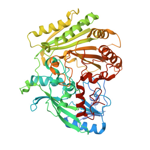
    Elucidation of natural product biosynthetic pathways provides important insights into the assembly of potent bioactive molecules, and expands access to unique enzymes able to selectively modify complex substrates. Here, we show full reconstitution, in vitro, of an unusual multi-step oxidative cascade for post-assembly-line tailoring of tirandamycin antibiotics. This pathway involves a remarkably versatile and iterative cytochrome P450 monooxygenase (TamI) and a flavin adenine dinucleotide-dependent oxidase (TamL), which act co-dependently through the repeated exchange of substrates. TamI hydroxylates tirandamycin C (TirC) to generate tirandamycin E (TirE), a previously unidentified tirandamycin intermediate. TirE is subsequently oxidized by TamL, giving rise to the ketone of tirandamycin D (TirD), after which a unique exchange back to TamI enables successive epoxidation and hydroxylation to afford, respectively, the final products tirandamycin A (TirA) and tirandamycin B (TirB). Ligand-free, substrate- and product-bound crystal structures of bicovalently flavinylated TamL oxidase reveal a likely mechanism for the C10 oxidation of TirE.


  • Organizational Affiliation
    • Life Sciences Institute and Department of Medicinal Chemistry, University of Michigan, Ann Arbor, Michigan 48109, USA.

Macromolecule Content 

  • Total Structure Weight: 238.47 kDa 
  • Atom Count: 17,755 
  • Modeled Residue Count: 1,962 
  • Deposited Residue Count: 2,120 
  • Unique protein chains: 1

Macromolecules

Find similar proteins by:|  3D Structure
Entity ID: 1
MoleculeChains  Sequence LengthOrganismDetailsImage
TAML
A, B, C, D
530Streptomyces sp. 307-9Mutation(s): 0 
UniProt
Find proteins for D3Y1I2 (Streptomyces sp. 307-9)
Explore D3Y1I2 
Go to UniProtKB:  D3Y1I2
Entity Groups
Sequence Clusters30% Identity50% Identity70% Identity90% Identity95% Identity100% Identity
UniProt GroupD3Y1I2
Sequence Annotations
Expand
Reference Sequence

Small Molecules

Ligands 6 Unique
IDChains Name / Formula / InChI Key2D Diagram3D Interactions
FAD

Query on FAD


Download:Ideal Coordinates CCD File
E [auth A],
J [auth B],
P [auth C],
W [auth D]
FLAVIN-ADENINE DINUCLEOTIDE
C27 H33 N9 O15 P2
VWWQXMAJTJZDQX-UYBVJOGSSA-N
TIR

Query on TIR


Download:Ideal Coordinates CCD File
F [auth A],
Q [auth C]
TIRANDAMYCIN E
C22 H29 N O6
ADEAEUKXVJOXIM-YNKBRHJSSA-N
TRK

Query on TRK


Download:Ideal Coordinates CCD File
K [auth B],
X [auth D]
TIRANDAMYCIN D
C22 H27 N O6
WORJTWSOUPGODS-HYAARBKISA-N
GOL

Query on GOL


Download:Ideal Coordinates CCD File
AA [auth D]
H [auth A]
M [auth B]
N [auth B]
S [auth C]
AA [auth D],
H [auth A],
M [auth B],
N [auth B],
S [auth C],
T [auth C],
Z [auth D]
GLYCEROL
C3 H8 O3
PEDCQBHIVMGVHV-UHFFFAOYSA-N
CL

Query on CL


Download:Ideal Coordinates CCD File
BA [auth D]
CA [auth D]
I [auth A]
O [auth B]
U [auth C]
BA [auth D],
CA [auth D],
I [auth A],
O [auth B],
U [auth C],
V [auth C]
CHLORIDE ION
Cl
VEXZGXHMUGYJMC-UHFFFAOYSA-M
MG

Query on MG


Download:Ideal Coordinates CCD File
G [auth A],
L [auth B],
R [auth C],
Y [auth D]
MAGNESIUM ION
Mg
JLVVSXFLKOJNIY-UHFFFAOYSA-N

Experimental Data & Validation

Experimental Data

  • Method: X-RAY DIFFRACTION
  • Resolution: 1.79 Å
  • R-Value Free:  0.219 (Depositor), 0.218 (DCC) 
  • R-Value Work:  0.146 (Depositor), 0.146 (DCC) 
  • R-Value Observed: 0.150 (Depositor) 
Space Group: P 1 21 1
Unit Cell:
Length ( Å )Angle ( ˚ )
a = 71.088α = 90
b = 106.171β = 94.63
c = 151.172γ = 90
Software Package:
Software NamePurpose
REFMACrefinement
MOSFLMdata reduction
SCALAdata scaling
PHASERphasing

Structure Validation

View Full Validation Report


Ligand Structure Quality Assessment 


Entry History 

Deposition Data

Revision History  (Full details and data files)

  • Version 1.0: 2011-06-29
    Type: Initial release
  • Version 1.1: 2011-08-03
    Changes: Database references, Non-polymer description, Version format compliance
  • Version 1.2: 2019-05-08
    Changes: Data collection, Derived calculations, Experimental preparation, Other
  • Version 1.3: 2023-12-20
    Changes: Data collection, Database references, Derived calculations, Other, Refinement description
  • Version 1.4: 2024-10-23
    Changes: Structure summary