2Y3A

Crystal structure of p110beta in complex with icSH2 of p85beta and the drug GDC-0941


Experimental Data Snapshot

  • Method: X-RAY DIFFRACTION
  • Resolution: 3.30 Å
  • R-Value Free: 0.296 
  • R-Value Work: 0.234 
  • R-Value Observed: 0.237 

wwPDB Validation   3D Report Full Report


Ligand Structure Quality Assessment 


This is version 1.4 of the entry. See complete history


Literature

Structure of Lipid Kinase P110Beta-P85Beta Elucidates an Unusual Sh2-Domain-Mediated Inhibitory Mechanism.

Zhang, X.Vadas, O.Perisic, O.Anderson, K.E.Clark, J.Hawkins, P.T.Stephens, L.R.Williams, R.L.

(2011) Mol Cell 41: 567

  • DOI: https://doi.org/10.1016/j.molcel.2011.01.026
  • Primary Citation of Related Structures:  
    2Y3A

  • PubMed Abstract: 

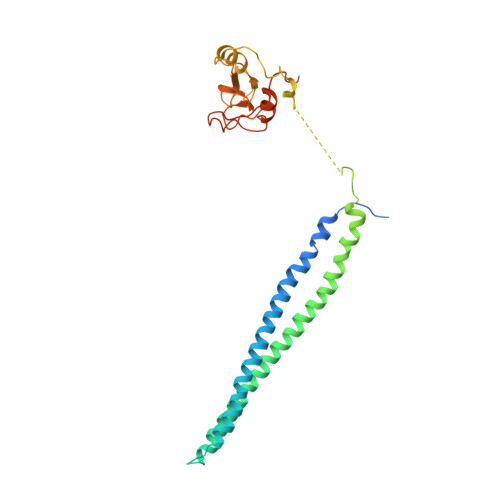
    Phosphoinositide 3-kinases (PI3Ks) are essential for cell growth, migration, and survival. The structure of a p110β/p85β complex identifies an inhibitory function for the C-terminal SH2 domain (cSH2) of the p85 regulatory subunit. Mutagenesis of a cSH2 contact residue activates downstream signaling in cells. This inhibitory contact ties up the C-terminal region of the p110β catalytic subunit, which is essential for lipid kinase activity. In vitro, p110β basal activity is tightly restrained by contacts with three p85 domains: the cSH2, nSH2, and iSH2. RTK phosphopeptides relieve inhibition by nSH2 and cSH2 using completely different mechanisms. The binding site for the RTK's pYXXM motif is exposed on the cSH2, requiring an extended RTK motif to reach and disrupt the inhibitory contact with p110β. This contrasts with the nSH2 where the pY-binding site itself forms the inhibitory contact. This establishes an unusual mechanism by which p85 SH2 domains contribute to RTK signaling specificities.


  • Organizational Affiliation

    Medical Research Council Laboratory of Molecular Biology, Hills Road, Cambridge CB2 0QH, UK.


Macromolecules
Find similar proteins by:  (by identity cutoff)  |  3D Structure
Entity ID: 1
MoleculeChains Sequence LengthOrganismDetailsImage
PHOSPHATIDYLINOSITOL-4,5-BISPHOSPHATE 3-KINASE CATALYTIC SUBUNIT BETA ISOFORM1,092Mus musculusMutation(s): 0 
EC: 2.7.1.173
UniProt & NIH Common Fund Data Resources
Find proteins for Q8BTI9 (Mus musculus)
Explore Q8BTI9 
Go to UniProtKB:  Q8BTI9
IMPC:  MGI:1922019
Entity Groups  
Sequence Clusters30% Identity50% Identity70% Identity90% Identity95% Identity100% Identity
UniProt GroupQ8BTI9
Sequence Annotations
Expand
  • Reference Sequence
Find similar proteins by:  (by identity cutoff)  |  3D Structure
Entity ID: 2
MoleculeChains Sequence LengthOrganismDetailsImage
PHOSPHATIDYLINOSITOL 3-KINASE REGULATORY SUBUNIT BETA302Mus musculusMutation(s): 0 
UniProt & NIH Common Fund Data Resources
Find proteins for O08908 (Mus musculus)
Explore O08908 
Go to UniProtKB:  O08908
IMPC:  MGI:1098772
Entity Groups  
Sequence Clusters30% Identity50% Identity70% Identity90% Identity95% Identity100% Identity
UniProt GroupO08908
Sequence Annotations
Expand
  • Reference Sequence
Small Molecules
Ligands 1 Unique
IDChains Name / Formula / InChI Key2D Diagram3D Interactions
GD9
Query on GD9

Download Ideal Coordinates CCD File 
C [auth A]2-(1H-indazol-4-yl)-6-{[4-(methylsulfonyl)piperazin-1-yl]methyl}-4-morpholin-4-yl-thieno[3,2-d]pyrimidine
C23 H27 N7 O3 S2
LHNIIDJUOCFXAP-UHFFFAOYSA-N
Binding Affinity Annotations 
IDSourceBinding Affinity
GD9 BindingDB:  2Y3A Ki: min: 70.2, max: 70.2 (nM) from 2 assay(s)
Kd: min: 2.1, max: 16 (nM) from 2 assay(s)
IC50: min: 3, max: 324 (nM) from 8 assay(s)
Experimental Data & Validation

Experimental Data

  • Method: X-RAY DIFFRACTION
  • Resolution: 3.30 Å
  • R-Value Free: 0.296 
  • R-Value Work: 0.234 
  • R-Value Observed: 0.237 
  • Space Group: P 65 2 2
Unit Cell:
Length ( Å )Angle ( ˚ )
a = 134.314α = 90
b = 134.314β = 90
c = 428.062γ = 120
Software Package:
Software NamePurpose
BUSTERrefinement
MOSFLMdata reduction
SCALAdata scaling
PHASERphasing

Structure Validation

View Full Validation Report


Ligand Structure Quality Assessment 


Entry History 

Deposition Data

Revision History  (Full details and data files)

  • Version 1.0: 2011-03-16
    Type: Initial release
  • Version 1.1: 2011-05-08
    Changes: Version format compliance
  • Version 1.2: 2011-07-13
    Changes: Version format compliance
  • Version 1.3: 2019-04-03
    Changes: Data collection, Other, Source and taxonomy
  • Version 1.4: 2023-12-20
    Changes: Data collection, Database references, Derived calculations, Other, Refinement description