


Methylphosphonate Adducts of Acetylcholinesterase Investigated by Time Correlated Single Photon Counting and X-Ray Crystallography
Akfur, C., Artursson, E., Ekstrom, F.To be published.
Experimental Data Snapshot
Starting Model: experimental
View more details
Entity ID: 1 | |||||
|---|---|---|---|---|---|
| Molecule | Chains | Sequence Length | Organism | Details | Image |
| ACETYLCHOLINESTERASE | 548 | Mus musculus | Mutation(s): 0 EC: 3.1.1.7 | 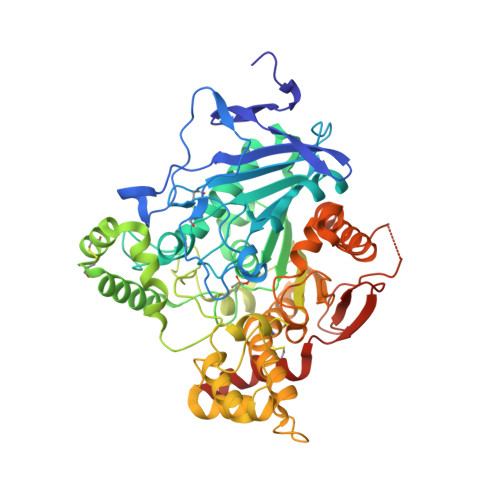 | |
UniProt & NIH Common Fund Data Resources | |||||
IMPC: MGI:87876 | |||||
Entity Groups | |||||
| Sequence Clusters | 30% Identity50% Identity70% Identity90% Identity95% Identity100% Identity | ||||
| UniProt Group | P21836 | ||||
Glycosylation | |||||
| Glycosylation Sites: 1 | Go to GlyGen: P21836-1 | ||||
Sequence AnnotationsExpand | |||||
Reference Sequence | |||||
| Ligands 5 Unique | |||||
|---|---|---|---|---|---|
| ID | Chains | Name / Formula / InChI Key | 2D Diagram | 3D Interactions | |
| P15 Download:Ideal Coordinates CCD File | H [auth A] | 2,5,8,11,14,17-HEXAOXANONADECAN-19-OL C13 H28 O7 FHHGCKHKTAJLOM-UHFFFAOYSA-N |  | ||
| NAG Download:Ideal Coordinates CCD File | C [auth A], D [auth A], J [auth B] | 2-acetamido-2-deoxy-beta-D-glucopyranose C8 H15 N O6 OVRNDRQMDRJTHS-FMDGEEDCSA-N |  | ||
| ME2 Download:Ideal Coordinates CCD File | G [auth A], L [auth B] | 1-ETHOXY-2-(2-METHOXYETHOXY)ETHANE C7 H16 O3 CNJRPYFBORAQAU-UHFFFAOYSA-N |  | ||
| PEG Download:Ideal Coordinates CCD File | E [auth A], F [auth A], I [auth A] | DI(HYDROXYETHYL)ETHER C4 H10 O3 MTHSVFCYNBDYFN-UHFFFAOYSA-N |  | ||
| ETX Download:Ideal Coordinates CCD File | K [auth B] | 2-ETHOXYETHANOL C4 H10 O2 ZNQVEEAIQZEUHB-UHFFFAOYSA-N |  | ||
| Modified Residues 1 Unique | |||||
|---|---|---|---|---|---|
| ID | Chains | Type | Formula | 2D Diagram | Parent |
| SGB Query on SGB | A, B | L-PEPTIDE LINKING | C7 H16 N O5 P |  | SER |
| Length ( Å ) | Angle ( ˚ ) |
|---|---|
| a = 79.488 | α = 90 |
| b = 111.643 | β = 90 |
| c = 227.026 | γ = 90 |
| Software Name | Purpose |
|---|---|
| PHENIX | refinement |
| XDS | data reduction |
| SCALA | data scaling |
| REFMAC | phasing |


