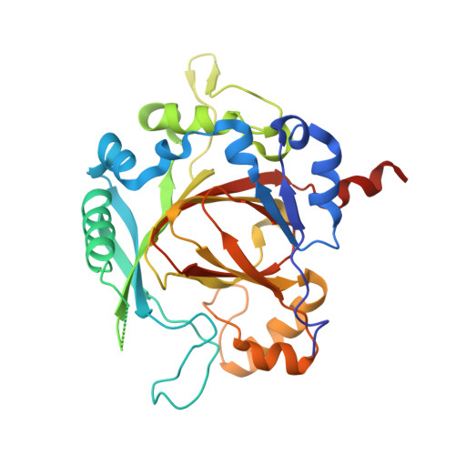
2XXZ | pdb_00002xxz

Crystal structure of the human JMJD3 jumonji domain


Experimental Data Snapshot

  • Method: X-RAY DIFFRACTION
  • Resolution: 1.80 Å
  • R-Value Free: 
    0.216 (Depositor), 0.245 (DCC) 
  • R-Value Work: 
    0.183 (Depositor), 0.213 (DCC) 
  • R-Value Observed: 
    0.185 (Depositor) 

wwPDB Validation 3D Report Full Report

Validation slider image for 2XXZ

Ligand Structure Quality Assessment 


This is version 1.3 of the entry. See complete history

Literature

Macromolecule Content 

  • Total Structure Weight: 76.26 kDa 
  • Atom Count: 5,082 
  • Modeled Residue Count: 578 
  • Deposited Residue Count: 664 
  • Unique protein chains: 1

Macromolecules

Find similar proteins by:|  3D Structure
Entity ID: 1
MoleculeChains  Sequence LengthOrganismDetailsImage
LYSINE-SPECIFIC DEMETHYLASE 6B
A, B
332Homo sapiensMutation(s): 0 
EC: 1.14.11 (PDB Primary Data), 1.14.11.68 (UniProt)
UniProt & NIH Common Fund Data Resources
Find proteins for O15054 (Homo sapiens)
Explore O15054 
Go to UniProtKB:  O15054
PHAROS:  O15054
GTEx:  ENSG00000132510 
Entity Groups
Sequence Clusters30% Identity50% Identity70% Identity90% Identity95% Identity100% Identity
UniProt GroupO15054
Sequence Annotations
Expand
Reference Sequence

Small Molecules

Ligands 4 Unique
IDChains Name / Formula / InChI Key2D Diagram3D Interactions
8XQ

Query on 8XQ


Download:Ideal Coordinates CCD File
J [auth A],
M [auth B]
8-hydroxyquinoline-5-carboxylic acid
C10 H7 N O3
JGRPKOGHYBAVMW-UHFFFAOYSA-N
EDO

Query on EDO


Download:Ideal Coordinates CCD File
F [auth A],
G [auth A],
H [auth A],
I [auth A],
L [auth B]
1,2-ETHANEDIOL
C2 H6 O2
LYCAIKOWRPUZTN-UHFFFAOYSA-N
NI

Query on NI


Download:Ideal Coordinates CCD File
C [auth A],
E [auth A],
K [auth B]
NICKEL (II) ION
Ni
VEQPNABPJHWNSG-UHFFFAOYSA-N
NA

Query on NA


Download:Ideal Coordinates CCD File
D [auth A]SODIUM ION
Na
FKNQFGJONOIPTF-UHFFFAOYSA-N
Binding Affinity Annotations 
IDSourceBinding Affinity
8XQ BindingDB:  2XXZ IC50: min: 100, max: 200 (nM) from 3 assay(s)

Experimental Data & Validation

Experimental Data

  • Method: X-RAY DIFFRACTION
  • Resolution: 1.80 Å
  • R-Value Free:  0.216 (Depositor), 0.245 (DCC) 
  • R-Value Work:  0.183 (Depositor), 0.213 (DCC) 
  • R-Value Observed: 0.185 (Depositor) 
Space Group: P 21 21 21
Unit Cell:
Length ( Å )Angle ( ˚ )
a = 56.757α = 90
b = 71.431β = 90
c = 159.698γ = 90
Software Package:
Software NamePurpose
REFMACrefinement
MOSFLMdata reduction
SCALAdata scaling

Structure Validation

View Full Validation Report


Ligand Structure Quality Assessment 


Entry History 

Revision History  (Full details and data files)

  • Version 1.0: 2010-11-24
    Type: Initial release
  • Version 1.1: 2015-01-14
    Changes: Non-polymer description, Version format compliance
  • Version 1.2: 2018-01-24
    Changes: Database references
  • Version 1.3: 2024-05-08
    Changes: Data collection, Database references, Derived calculations, Other