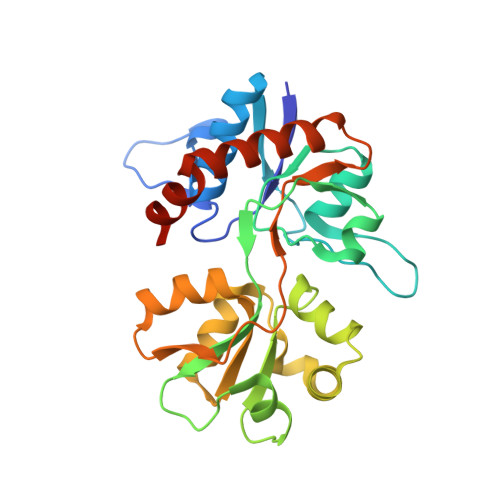
Conformational Flexibility of the Ligand-Binding Domain Dimer in Kainate Receptor Gating and Desensitization
Nayeem, N., Mayans, O., Green, T.(2011) J Neurosci 31: 2916
- PubMed: 21414913
- DOI: https://doi.org/10.1523/JNEUROSCI.4771-10.2011
- Primary Citation of Related Structures:
2XXR, 2XXT, 2XXU, 2XXV, 2XXW, 2XXX, 2XXY - PubMed Abstract:
AMPA- and kainate (KA)-selective ionotropic glutamate receptors (iGluRs) respond to agonist by opening (gating), then closing (desensitizing) in quick succession. Gating has been linked to agonist-induced changes within the ligand-binding domain (LBD), and desensitization to rearrangement of a dimer formed by neighboring LBDs. To explore the role of dimer conformation in both gating and desensitization, we compared the conformational effects of two kainate receptor mutants. The first, GluK2-D776K, blocks desensitization of macroscopic current responses ("macroscopic desensitization"). The second, GluK2-M770K, accelerates macroscopic desensitization and eliminates the effects of external ions on channel kinetics. Using structures determined by x-ray crystallography, we found that in both mutants the introduced lysines act as tethered cations, displacing sodium ions from their binding sites within the dimer interface. This results in new inter- and intra-protomer contacts in D776K and M770K respectively, explaining the effects of these mutations on dimer stability and desensitization kinetics. Further, chloride binding was unaffected by the M770K mutation, even though binding of sodium ions has been proposed to promote dimer stability by stabilizing anion binding. This suggests sodium binding may affect receptor function more directly than currently supposed. Notably, we also observed a ligand-specific shift in dimer conformation when comparing LBD dimers in complex with glutamate or the partial agonist KA, revealing a previously unidentified role for dimer orientation in iGluR gating.
- Department of Pharmacology, University of Liverpool, Liverpool L69 3BX, United Kingdom.
Organizational Affiliation:


