Inhibitors of the Tyrosine Kinase Ephb4. Part 4: Discovery and Optimization of a Benzylic Alcohol Series.
Barlaam, B., Ducray, R., Lambert-Van Der Brempt, C., Ple, P., Bardelle, C., Brooks, N., Coleman, T., Cross, D., Kettle, J.G., Read, J.(2011) Bioorg Med Chem Lett 21: 2207
- PubMed: 21441027 Search on PubMed
- DOI: https://doi.org/10.1016/j.bmcl.2011.03.009
- Primary Citation Related Structures:
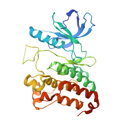
2XVD - PubMed Abstract:
Optimization of our bis-anilino-pyrimidine series of EphB4 kinase inhibitors led to the discovery of compound 12 which incorporates a key m-hydroxymethylene group on the C4 aniline. 12 displays a good kinase selectivity profile, good physical properties and pharmacokinetic parameters, suggesting it is a suitable candidate to investigate the therapeutic potential of EphB4 kinase inhibitors.
- AstraZeneca, Centre de Recherches, Z.I. la Pompelle, BP1050, 51689 Reims, Cedex 2, France.
Organizational Affiliation: