2XV1 | pdb_00002xv1

Crystal structure of the triscatecholate siderophore binding protein FeuA from Bacillus subtilis complexed with Ferric MECAM


Experimental Data Snapshot

  • Method: X-RAY DIFFRACTION
  • Resolution: 2.15 Å
  • R-Value Free: 
    0.237 (Depositor), 0.241 (DCC) 
  • R-Value Work: 
    0.178 (Depositor), 0.182 (DCC) 
  • R-Value Observed: 
    0.183 (Depositor) 

Starting Model: experimental
View more details

wwPDB Validation 3D Report Full Report

Validation slider image for 2XV1

Ligand Structure Quality Assessment 


This is version 1.2 of the entry. See complete history

Literature

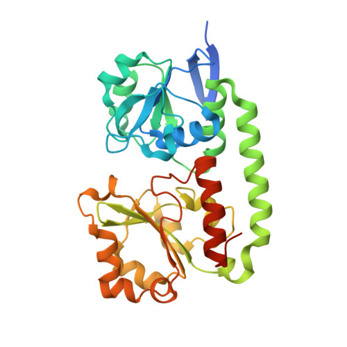
The Siderophore Binding Protein Feua Shows Limited Promiscuity Toward Exogenous Triscatecholates

Peuckert, F.Ramos-Vega, A.L.Miethke, M.Schwoerer, C.J.Albrecht, A.G.Oberthuer, M.Marahiel, M.A.

(2011) Chem Biol 18: 907

  • DOI: https://doi.org/10.1016/j.chembiol.2011.05.006
  • Primary Citation Related Structures: 
    2XUZ, 2XV1

  • PubMed Abstract: 

    Iron acquisition by siderophores is crucial for survival and virulence of many microorganisms. Here, we investigated the binding of the exogenous siderophore ferric enterobactin and the synthetic siderophore mimic ferric mecam by the triscatecholate binding protein FeuA from Bacillus subtilis at the atomic level. The structural complexes provide molecular insights into the capture mechanism of FeuA for exogenous and synthetic siderophores. The protein-ligand complexes show an exclusive acceptance of Λ-stereoconfigured substrates. Ligand-induced cross-bridging of the complexes was not observed, revealing a different thermodynamic behavior especially of the ferric mecam substrate, which was previously shown to dimerize with the enterobactin binding protein CeuE. The nearly identical overall domain movement of FeuA upon binding of ferric enterobactin or ferric mecam compared with endogenously derived ferric bacillibactin implies the importance of the conserved domain rearrangement for recognition by the transmembrane permease FeuBC, for which the conserved FeuA residues E90 and E221 were proved to be essential.


  • Organizational Affiliation
    • Department of Chemistry, Biochemistry, Philipps-University Marburg, Hans-Meerwein-Straße, D-35032 Marburg, Germany.

Macromolecule Content 

  • Total Structure Weight: 35.78 kDa 
  • Atom Count: 2,314 
  • Modeled Residue Count: 283 
  • Deposited Residue Count: 311 
  • Unique protein chains: 1

Macromolecules

Find similar proteins by:|  3D Structure
Entity ID: 1
MoleculeChains  Sequence LengthOrganismDetailsImage
IRON-UPTAKE SYSTEM-BINDING PROTEIN311Bacillus subtilisMutation(s): 0 
UniProt
Find proteins for P40409 (Bacillus subtilis (strain 168))
Explore P40409 
Go to UniProtKB:  P40409
Entity Groups
Sequence Clusters30% Identity50% Identity70% Identity90% Identity95% Identity100% Identity
UniProt GroupP40409
Sequence Annotations
Expand
Reference Sequence

Small Molecules

Ligands 4 Unique
IDChains Name / Formula / InChI Key2D Diagram3D Interactions
ECA

Query on ECA


Download:Ideal Coordinates CCD File
B [auth A]N,N',N''-[BENZENE-1,3,5-TRIYLTRIS(METHYLENE)]TRIS(2,3-DIHYDROXYBENZAMIDE)
C30 H27 N3 O9
GCTFIRZGPIUOAK-UHFFFAOYSA-N
PEG

Query on PEG


Download:Ideal Coordinates CCD File
F [auth A],
G [auth A]
DI(HYDROXYETHYL)ETHER
C4 H10 O3
MTHSVFCYNBDYFN-UHFFFAOYSA-N
PO4

Query on PO4


Download:Ideal Coordinates CCD File
D [auth A],
E [auth A]
PHOSPHATE ION
O4 P
NBIIXXVUZAFLBC-UHFFFAOYSA-K
FE

Query on FE


Download:Ideal Coordinates CCD File
C [auth A]FE (III) ION
Fe
VTLYFUHAOXGGBS-UHFFFAOYSA-N

Experimental Data & Validation

Experimental Data

  • Method: X-RAY DIFFRACTION
  • Resolution: 2.15 Å
  • R-Value Free:  0.237 (Depositor), 0.241 (DCC) 
  • R-Value Work:  0.178 (Depositor), 0.182 (DCC) 
  • R-Value Observed: 0.183 (Depositor) 
Space Group: P 1 21 1
Unit Cell:
Length ( Å )Angle ( ˚ )
a = 39.96α = 90
b = 63.44β = 110.27
c = 56.21γ = 90
Software Package:
Software NamePurpose
REFMACrefinement
XDSdata reduction
XSCALEdata scaling
PHASERphasing

Structure Validation

View Full Validation Report


Ligand Structure Quality Assessment 


Entry History 

Deposition Data

Revision History  (Full details and data files)

  • Version 1.0: 2011-08-10
    Type: Initial release
  • Version 1.1: 2012-03-28
    Changes: Other
  • Version 1.2: 2023-12-20
    Changes: Data collection, Database references, Derived calculations, Other, Refinement description