2XTZ | pdb_00002xtz

Crystal structure of the G alpha protein AtGPA1 from Arabidopsis thaliana


Experimental Data Snapshot

  • Method: X-RAY DIFFRACTION
  • Resolution: 2.34 Å
  • R-Value Free: 
    0.254 (Depositor), 0.247 (DCC) 
  • R-Value Work: 
    0.211 (Depositor), 0.204 (DCC) 
  • R-Value Observed: 
    0.212 (Depositor) 

Starting Model: experimental
View more details

wwPDB Validation 3D Report Full Report

Validation slider image for 2XTZ

Ligand Structure Quality Assessment 


This is version 1.6 of the entry. See complete history

Literature

The Crystal Structure of a Self-Activating G Protein Alpha Subunit Reveals its Distinct Mechanism of Signal Initiation

Jones, J.C.Duffy, J.W.Machius, M.Temple, B.R.S.Dohlman, H.G.Jones, A.M.

(2011) Sci Signal 159: RA8

  • DOI: https://doi.org/10.1126/scisignal.2001446
  • Primary Citation Related Structures: 
    2XTZ

  • PubMed Abstract: 

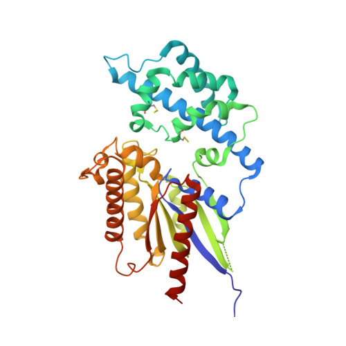
    In animals, heterotrimeric guanine nucleotide-binding protein (G protein) signaling is initiated by G protein-coupled receptors (GPCRs), which activate G protein α subunits; however, the plant Arabidopsis thaliana lacks canonical GPCRs, and its G protein α subunit (AtGPA1) is self-activating. To investigate how AtGPA1 becomes activated, we determined its crystal structure. AtGPA1 is structurally similar to animal G protein α subunits, but our crystallographic and biophysical studies revealed that it had distinct properties. Notably, the helical domain of AtGPA1 displayed pronounced intrinsic disorder and a tendency to disengage from the Ras domain of the protein. Domain substitution experiments showed that the helical domain of AtGPA1 was necessary for self-activation and sufficient to confer self-activation to an animal G protein α subunit. These findings reveal the structural basis for a mechanism for G protein activation in Arabidopsis that is distinct from the well-established mechanism found in animals.


  • Organizational Affiliation
    • Department of Biochemistry and Biophysics, University of North Carolina, Chapel Hill, NC 27599, USA.

Macromolecule Content 

  • Total Structure Weight: 125.54 kDa 
  • Atom Count: 7,909 
  • Modeled Residue Count: 904 
  • Deposited Residue Count: 1,062 
  • Unique protein chains: 1

Macromolecules

Find similar proteins by:|  3D Structure
Entity ID: 1
MoleculeChains  Sequence LengthOrganismDetailsImage
GUANINE NUCLEOTIDE-BINDING PROTEIN ALPHA-1 SUBUNIT
A, B, C
354Arabidopsis thalianaMutation(s): 0 
EC: 3.6.5.1
UniProt
Find proteins for P18064 (Arabidopsis thaliana)
Explore P18064 
Go to UniProtKB:  P18064
Entity Groups
Sequence Clusters30% Identity50% Identity70% Identity90% Identity95% Identity100% Identity
UniProt GroupP18064
Sequence Annotations
Expand
Reference Sequence

Small Molecules

Ligands 4 Unique
IDChains Name / Formula / InChI Key2D Diagram3D Interactions
GSP

Query on GSP


Download:Ideal Coordinates CCD File
E [auth A],
I [auth B],
N [auth C]
5'-GUANOSINE-DIPHOSPHATE-MONOTHIOPHOSPHATE
C10 H16 N5 O13 P3 S
XOFLBQFBSOEHOG-UUOKFMHZSA-N
SO4

Query on SO4


Download:Ideal Coordinates CCD File
G [auth A],
K [auth B],
O [auth C],
P [auth C],
Q [auth C]
SULFATE ION
O4 S
QAOWNCQODCNURD-UHFFFAOYSA-L
CL

Query on CL


Download:Ideal Coordinates CCD File
F [auth A],
J [auth B],
L [auth B],
R [auth C]
CHLORIDE ION
Cl
VEXZGXHMUGYJMC-UHFFFAOYSA-M
MG

Query on MG


Download:Ideal Coordinates CCD File
D [auth A],
H [auth B],
M [auth C]
MAGNESIUM ION
Mg
JLVVSXFLKOJNIY-UHFFFAOYSA-N
Modified Residues  1 Unique
IDChains TypeFormula2D DiagramParent
CSS
Query on CSS
A, B, C
L-PEPTIDE LINKINGC3 H7 N O2 S2CYS

Experimental Data & Validation

Experimental Data

  • Method: X-RAY DIFFRACTION
  • Resolution: 2.34 Å
  • R-Value Free:  0.254 (Depositor), 0.247 (DCC) 
  • R-Value Work:  0.211 (Depositor), 0.204 (DCC) 
  • R-Value Observed: 0.212 (Depositor) 
Space Group: P 21 21 21
Unit Cell:
Length ( Å )Angle ( ˚ )
a = 67.13α = 90
b = 119.347β = 90
c = 161.725γ = 90
Software Package:
Software NamePurpose
REFMACrefinement
HKL-2000data reduction
HKL-2000data scaling
PHASERphasing

Structure Validation

View Full Validation Report


Ligand Structure Quality Assessment 


Entry History 

Deposition Data

Revision History  (Full details and data files)

  • Version 1.0: 2011-02-16
    Type: Initial release
  • Version 1.1: 2011-05-08
    Changes: Version format compliance
  • Version 1.2: 2011-07-13
    Changes: Version format compliance
  • Version 1.3: 2019-04-03
    Changes: Data collection, Derived calculations, Experimental preparation, Other
  • Version 1.4: 2019-05-15
    Changes: Data collection, Experimental preparation
  • Version 1.5: 2023-12-20
    Changes: Data collection, Database references, Derived calculations, Other, Refinement description
  • Version 1.6: 2024-11-13
    Changes: Structure summary