2XTE | pdb_00002xte

Structure of the TBL1 tetramerisation domain


Experimental Data Snapshot

  • Method: X-RAY DIFFRACTION
  • Resolution: 3.90 Å
  • R-Value Free: 
    0.302 (Depositor), 0.313 (DCC) 
  • R-Value Work: 
    0.273 (Depositor), 0.276 (DCC) 
  • R-Value Observed: 
    0.273 (Depositor) 

Starting Model: experimental
View more details

wwPDB Validation 3D Report Full Report

Validation slider image for 2XTE

This is version 1.3 of the entry. See complete history

Literature

Structural Basis for the Assembly of the Smrt/Ncor Core Transcriptional Repression Machinery.

Oberoi, J.Fairall, L.Watson, P.J.Yang, J.C.Czimmerer, Z.Kampmann, T.Goult, B.T.Greenwood, J.A.Gooch, J.T.Kallenberger, B.C.Nagy, L.Neuhaus, D.Schwabe, J.W.R.

(2011) Nat Struct Mol Biol 18: 177

  • DOI: https://doi.org/10.1038/nsmb.1983
  • Primary Citation Related Structures: 
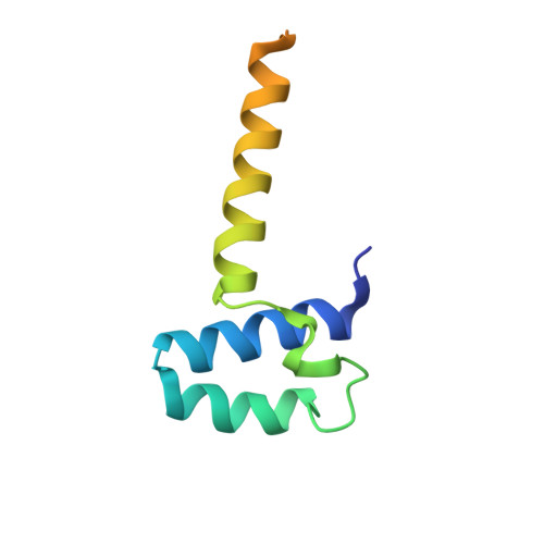
    2L5G, 2XTC, 2XTD, 2XTE

  • PubMed Abstract: 

    Eukaryotic transcriptional repressors function by recruiting large coregulatory complexes that target histone deacetylase enzymes to gene promoters and enhancers. Transcriptional repression complexes, assembled by the corepressor NCoR and its homolog SMRT, are crucial in many processes, including development and metabolic physiology. The core repression complex involves the recruitment of three proteins, HDAC3, GPS2 and TBL1, to a highly conserved repression domain within SMRT and NCoR. We have used structural and functional approaches to gain insight into the architecture and biological role of this complex. We report the crystal structure of the tetrameric oligomerization domain of TBL1, which interacts with both SMRT and GPS2, and the NMR structure of the interface complex between GPS2 and SMRT. These structures, together with computational docking, mutagenesis and functional assays, reveal the assembly mechanism and stoichiometry of the corepressor complex.


  • Organizational Affiliation
    • Henry Wellcome Laboratories of Structural Biology, Department of Biochemistry, University of Leicester, Leicester, UK.

Macromolecule Content 

  • Total Structure Weight: 118.09 kDa 
  • Atom Count: 6,036 
  • Modeled Residue Count: 792 
  • Deposited Residue Count: 1,080 
  • Unique protein chains: 1

Macromolecules

Find similar proteins by:|  3D Structure
Entity ID: 1
MoleculeChains  Sequence LengthOrganismDetailsImage
F-BOX-LIKE/WD REPEAT-CONTAINING PROTEIN TBL1X
A, B, C, D, E
A, B, C, D, E, F, G, H, I, J, K, L
90Homo sapiensMutation(s): 0 
UniProt & NIH Common Fund Data Resources
Find proteins for O60907 (Homo sapiens)
Explore O60907 
Go to UniProtKB:  O60907
PHAROS:  O60907
GTEx:  ENSG00000101849 
Entity Groups
Sequence Clusters30% Identity50% Identity70% Identity90% Identity95% Identity100% Identity
UniProt GroupO60907
Sequence Annotations
Expand
Reference Sequence

Experimental Data & Validation

Experimental Data

  • Method: X-RAY DIFFRACTION
  • Resolution: 3.90 Å
  • R-Value Free:  0.302 (Depositor), 0.313 (DCC) 
  • R-Value Work:  0.273 (Depositor), 0.276 (DCC) 
  • R-Value Observed: 0.273 (Depositor) 
Space Group: P 21 21 21
Unit Cell:
Length ( Å )Angle ( ˚ )
a = 88.02α = 90
b = 150.19β = 90
c = 164.83γ = 90
Software Package:
Software NamePurpose
CNSrefinement
MOSFLMdata reduction
SCALAdata scaling
PHASERphasing

Structure Validation

View Full Validation Report


Entry History 

Deposition Data

Revision History  (Full details and data files)

  • Version 1.0: 2011-01-19
    Type: Initial release
  • Version 1.1: 2011-05-08
    Changes: Version format compliance
  • Version 1.2: 2011-07-13
    Changes: Version format compliance
  • Version 1.3: 2023-12-20
    Changes: Data collection, Database references, Other, Refinement description