2XQB | pdb_00002xqb

Crystal Structure of anti-IL-15 Antibody in Complex with human IL-15


Experimental Data Snapshot

  • Method: X-RAY DIFFRACTION
  • Resolution: 2.60 Å
  • R-Value Free: 
    0.318 (Depositor) 
  • R-Value Work: 
    0.244 (Depositor), 0.258 (DCC) 
  • R-Value Observed: 
    0.248 (Depositor) 

Starting Model: experimental
View more details

wwPDB Validation 3D Report Full Report

Validation slider image for 2XQB

This is version 1.5 of the entry. See complete history

Literature

Engineering a High Affinity Anti-Il-15 Antibody: Crystal Structure Reveals an Alpha-Helix in Vh Cdr3 as Key Component of Paratope.

Lowe, D.C.Gerhardt, S.Ward, A.Hargreaves, D.Anderson, M.Ferraro, F.Pauptit, R.A.Pattison, D.V.Buchanan, C.Popovic, B.Finch, D.K.Wilkinson, T.Sleeman, M.Vaughan, T.J.Mallinder, P.R.

(2011) J Mol Biology 406: 160

  • DOI: https://doi.org/10.1016/j.jmb.2010.12.017
  • Primary Citation Related Structures: 
    2XQB

  • PubMed Abstract: 

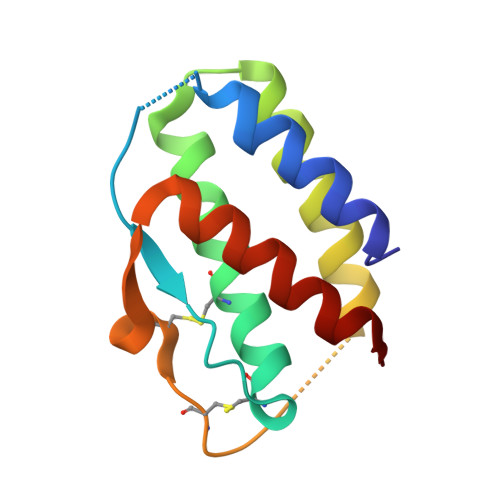
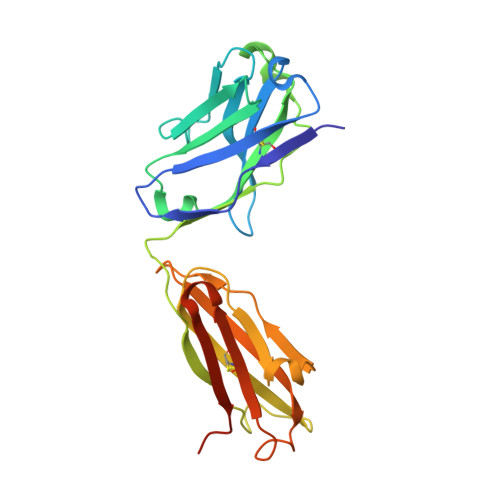
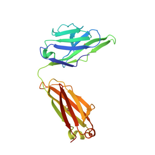
    Interleukin (IL) 15 is an inflammatory cytokine that plays an essential role in the activation, proliferation, and maintenance of specific natural killer cell and T-cell populations, and has been implicated as a mediator of inflammatory diseases. An anti-IL-15 antibody that blocked IL-15-dependent cellular responses was isolated by phage display and optimised via mutagenesis of the third complementarity-determining regions (CDRs) of variable heavy (VH) and variable light chains. Entire repertoires of improved variants were recombined with each other to explore the maximum potential sequence space. DISC0280, the most potent antibody isolated using this comprehensive strategy, exhibits a 228-fold increase in affinity and a striking 40,000-fold increase in cellular potency compared to its parent. Such a wholesale recombination strategy therefore represents a useful method for exploiting synergistic potency gains as part of future antibody engineering efforts. The crystal structure of DISC0280 Fab (fragment antigen binding), in complex with human IL-15, was determined in order to map the structural epitope and paratope. The most remarkable feature revealed lies within the paratope and is a novel six-amino-acid α-helix that sits within the VH CDR3 loop at the center of the antigen binding site. This is the first report to describe an α-helix as a principal component of a naturally derived VH CDR3 following affinity maturation.


  • Organizational Affiliation
    • MedImmune Ltd., Milstein Building, Granta Park, Cambridge CB1 6GH, UK. lowed@medimmune.com

Macromolecule Content 

  • Total Structure Weight: 61.38 kDa 
  • Atom Count: 4,063 
  • Modeled Residue Count: 534 
  • Deposited Residue Count: 561 
  • Unique protein chains: 3

Macromolecules

Find similar proteins by:|  3D Structure
Entity ID: 1
MoleculeChains  Sequence LengthOrganismDetailsImage
INTERLEUKIN 15114Homo sapiensMutation(s): 0 
UniProt & NIH Common Fund Data Resources
Find proteins for P40933 (Homo sapiens)
Explore P40933 
Go to UniProtKB:  P40933
PHAROS:  P40933
GTEx:  ENSG00000164136 
Entity Groups
Sequence Clusters30% Identity50% Identity70% Identity90% Identity95% Identity100% Identity
UniProt GroupP40933
Sequence Annotations
Expand
Reference Sequence
Find similar proteins by:|  3D Structure
Entity ID: 2
MoleculeChains  Sequence LengthOrganismDetailsImage
ANTI-IL-15 ANTIBODYB [auth H]236Homo sapiensMutation(s): 0 
Find similar proteins by:|  3D Structure
Entity ID: 3
MoleculeChains  Sequence LengthOrganismDetailsImage
ANTI-IL-15 ANTIBODYC [auth L]211Homo sapiensMutation(s): 0 

Experimental Data & Validation

Experimental Data

  • Method: X-RAY DIFFRACTION
  • Resolution: 2.60 Å
  • R-Value Free:  0.318 (Depositor) 
  • R-Value Work:  0.244 (Depositor), 0.258 (DCC) 
  • R-Value Observed: 0.248 (Depositor) 
Space Group: C 1 2 1
Unit Cell:
Length ( Å )Angle ( ˚ )
a = 185.186α = 90
b = 43.754β = 95.95
c = 70.087γ = 90
Software Package:
Software NamePurpose
REFMACrefinement
d*TREKdata reduction
d*TREKdata scaling
MOLREPphasing

Structure Validation

View Full Validation Report


Entry History 

Revision History  (Full details and data files)

  • Version 1.0: 2010-12-29
    Type: Initial release
  • Version 1.1: 2011-05-08
    Changes: Version format compliance
  • Version 1.2: 2011-07-13
    Changes: Version format compliance
  • Version 1.3: 2018-04-04
    Changes: Data collection, Source and taxonomy
  • Version 1.4: 2023-12-20
    Changes: Data collection, Database references, Derived calculations, Other, Refinement description
  • Version 1.5: 2024-10-23
    Changes: Structure summary