2XNX

BC1 fragment of streptococcal M1 protein in complex with human fibrinogen


Experimental Data Snapshot

  • Method: X-RAY DIFFRACTION
  • Resolution: 3.30 Å
  • R-Value Free: 0.325 
  • R-Value Work: 0.277 
  • R-Value Observed: 0.279 

wwPDB Validation   3D Report Full Report


This is version 1.2 of the entry. See complete history


Literature

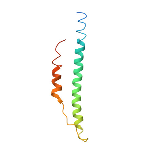
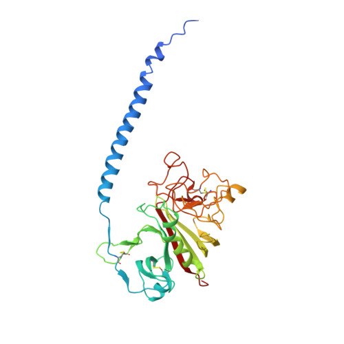
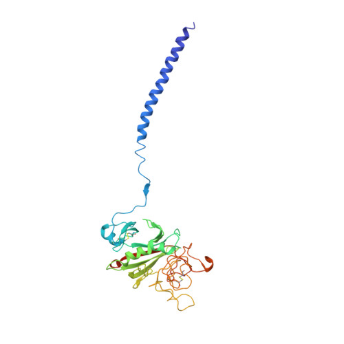
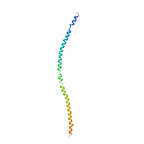
Streptococcal M1 Protein Constructs a Pathological Host Fibrinogen Network

Macheboeuf, P.Buffalo, C.Fu, C.Y.Zinkernagel, A.S.Cole, J.N.Johnson, J.E.Nizet, V.Nizet, V.Ghosh, P.

(2011) Nature 472: 64

  • DOI: https://doi.org/10.1038/nature09967
  • Primary Citation of Related Structures:  
    2XNX, 2XNY

  • PubMed Abstract: 
  • M1 protein, a major virulence factor of the leading invasive strain of group A Streptococcus, is sufficient to induce toxic-shock-like vascular leakage and tissue injury. These events are triggered by the formation of a complex between M1 and fibrinogen that, unlike M1 or fibrinogen alone, leads to neutrophil activation ...

    M1 protein, a major virulence factor of the leading invasive strain of group A Streptococcus, is sufficient to induce toxic-shock-like vascular leakage and tissue injury. These events are triggered by the formation of a complex between M1 and fibrinogen that, unlike M1 or fibrinogen alone, leads to neutrophil activation. Here we provide a structural explanation for the pathological properties of the complex formed between streptococcal M1 and human fibrinogen. A conformationally dynamic coiled-coil dimer of M1 was found to organize four fibrinogen molecules into a specific cross-like pattern. This pattern supported the construction of a supramolecular network that was required for neutrophil activation but was distinct from a fibrin clot. Disruption of this network into other supramolecular assemblies was not tolerated. These results have bearing on the pathophysiology of streptococcal toxic shock.


    Organizational Affiliation

    Department of Chemistry and Biochemistry, University of California, San Diego, La Jolla, California 92093, USA.


Macromolecules
Find similar proteins by:  (by identity cutoff)  |  3D Structure
Entity ID: 1
MoleculeChainsSequence LengthOrganismDetailsImage
FIBRINOGEN ALPHA CHAINA, D, G, J87Homo sapiensMutation(s): 0 
Gene Names: FGA
UniProt & NIH Common Fund Data Resources
Find proteins for P02671 (Homo sapiens)
Explore P02671 
Go to UniProtKB:  P02671
PHAROS:  P02671
Entity Groups  
Sequence Clusters30% Identity50% Identity70% Identity90% Identity95% Identity100% Identity
UniProt GroupP02671
Protein Feature View
Expand
  • Reference Sequence
Find similar proteins by:  (by identity cutoff)  |  3D Structure
Entity ID: 2
MoleculeChainsSequence LengthOrganismDetailsImage
FIBRINOGEN BETA CHAINB, E, H, K328Homo sapiensMutation(s): 0 
Gene Names: FGB
UniProt & NIH Common Fund Data Resources
Find proteins for P02675 (Homo sapiens)
Explore P02675 
Go to UniProtKB:  P02675
PHAROS:  P02675
Entity Groups  
Sequence Clusters30% Identity50% Identity70% Identity90% Identity95% Identity100% Identity
UniProt GroupP02675
Protein Feature View
Expand
  • Reference Sequence
Find similar proteins by:  (by identity cutoff)  |  3D Structure
Entity ID: 3
MoleculeChainsSequence LengthOrganismDetailsImage
FIBRINOGEN GAMMA CHAINC, F, I, L319Homo sapiensMutation(s): 0 
Gene Names: FGGPRO2061
UniProt & NIH Common Fund Data Resources
Find proteins for P02679 (Homo sapiens)
Explore P02679 
Go to UniProtKB:  P02679
PHAROS:  P02679
Entity Groups  
Sequence Clusters30% Identity50% Identity70% Identity90% Identity95% Identity100% Identity
UniProt GroupP02679
Protein Feature View
Expand
  • Reference Sequence
Find similar proteins by:  (by identity cutoff)  |  3D Structure
Entity ID: 4
MoleculeChainsSequence LengthOrganismDetailsImage
M PROTEINM, N146Streptococcus pyogenesMutation(s): 0 
Entity Groups  
Sequence Clusters30% Identity50% Identity70% Identity90% Identity95% Identity100% Identity
Protein Feature View
Expand
  • Reference Sequence
Experimental Data & Validation

Experimental Data

  • Method: X-RAY DIFFRACTION
  • Resolution: 3.30 Å
  • R-Value Free: 0.325 
  • R-Value Work: 0.277 
  • R-Value Observed: 0.279 
  • Space Group: P 1 21 1
Unit Cell:
Length ( Å )Angle ( ˚ )
a = 112.723α = 90
b = 216.866β = 102.54
c = 140.807γ = 90
Software Package:
Software NamePurpose
REFMACrefinement
MOSFLMdata reduction
SCALAdata scaling
PHASERphasing

Structure Validation

View Full Validation Report


Entry History 

Deposition Data

Revision History  (Full details and data files)

  • Version 1.0: 2011-04-13
    Type: Initial release
  • Version 1.1: 2011-05-08
    Changes: Version format compliance
  • Version 1.2: 2011-07-13
    Changes: Version format compliance