2XNG | pdb_00002xng

Structure of Aurora-A bound to a selective imidazopyrazine inhibitor


Experimental Data Snapshot

  • Method: X-RAY DIFFRACTION
  • Resolution: 2.60 Å
  • R-Value Free: 
    0.271 (Depositor), 0.262 (DCC) 
  • R-Value Work: 
    0.221 (Depositor), 0.216 (DCC) 
  • R-Value Observed: 
    0.224 (Depositor) 

Starting Model: experimental
View more details

wwPDB Validation 3D Report Full Report

Validation slider image for 2XNG

Ligand Structure Quality Assessment 


This is version 1.4 of the entry. See complete history

Literature

Structure-based design of imidazo[1,2-a]pyrazine derivatives as selective inhibitors of Aurora-A kinase in cells.

Bouloc, N.Large, J.M.Kosmopoulou, M.Sun, C.Faisal, A.Matteucci, M.Reynisson, J.Brown, N.Atrash, B.Blagg, J.McDonald, E.Linardopoulos, S.Bayliss, R.Bavetsias, V.

(2010) Bioorg Med Chem Lett 20: 5988-5993

  • DOI: https://doi.org/10.1016/j.bmcl.2010.08.091
  • Primary Citation Related Structures: 
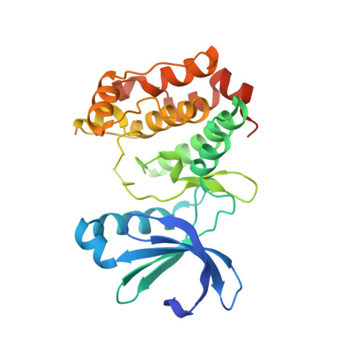
    2XNE, 2XNG

  • PubMed Abstract: 

    Co-crystallisation of the imidazo[1,2-a]pyrazine derivative 15 (3-chloro-N-(4-morpholinophenyl)-6-(pyridin-3-yl)imidazo[1,2-a]pyrazin-8-amine) with Aurora-A provided an insight into the interactions of this class of compound with Aurora kinases. This led to the design and synthesis of potent Aurora-A inhibitors demonstrating up to 70-fold selectivity in cell-based Aurora kinase pharmacodynamic biomarker assays.


  • Organizational Affiliation
    • Cancer Research UK Cancer Therapeutics Unit, The Institute of Cancer Research, Sutton, Surrey, United Kingdom.

Macromolecule Content 

  • Total Structure Weight: 33.33 kDa 
  • Atom Count: 2,036 
  • Modeled Residue Count: 254 
  • Deposited Residue Count: 283 
  • Unique protein chains: 1

Macromolecules

Find similar proteins by:|  3D Structure
Entity ID: 1
MoleculeChains  Sequence LengthOrganismDetailsImage
SERINE/THREONINE-PROTEIN KINASE 6283Homo sapiensMutation(s): 0 
EC: 2.7.11.1
UniProt & NIH Common Fund Data Resources
Find proteins for O14965 (Homo sapiens)
Explore O14965 
Go to UniProtKB:  O14965
PHAROS:  O14965
GTEx:  ENSG00000087586 
Entity Groups
Sequence Clusters30% Identity50% Identity70% Identity90% Identity95% Identity100% Identity
UniProt GroupO14965
Sequence Annotations
Expand
Reference Sequence

Small Molecules

Ligands 1 Unique
IDChains Name / Formula / InChI Key2D Diagram3D Interactions
A0H

Query on A0H


Download:Ideal Coordinates CCD File
B [auth A]N-(3-{3-chloro-8-[(4-morpholin-4-ylphenyl)amino]imidazo[1,2-a]pyrazin-6-yl}benzyl)methanesulfonamide
C24 H25 Cl N6 O3 S
BHSRVQWRAVCUOG-UHFFFAOYSA-N

Experimental Data & Validation

Experimental Data

  • Method: X-RAY DIFFRACTION
  • Resolution: 2.60 Å
  • R-Value Free:  0.271 (Depositor), 0.262 (DCC) 
  • R-Value Work:  0.221 (Depositor), 0.216 (DCC) 
  • R-Value Observed: 0.224 (Depositor) 
Space Group: P 61 2 2
Unit Cell:
Length ( Å )Angle ( ˚ )
a = 82.096α = 90
b = 82.096β = 90
c = 171.628γ = 120
Software Package:
Software NamePurpose
PHENIXrefinement

Structure Validation

View Full Validation Report


Ligand Structure Quality Assessment 


Entry History 

Deposition Data

Revision History  (Full details and data files)

  • Version 1.0: 2010-09-22
    Type: Initial release
  • Version 1.1: 2011-05-08
    Changes: Version format compliance
  • Version 1.2: 2011-07-13
    Changes: Version format compliance
  • Version 1.3: 2018-02-28
    Changes: Database references
  • Version 1.4: 2023-12-20
    Changes: Data collection, Database references, Derived calculations, Other, Refinement description