


Binding Diversity of Pioglitazone by Peroxisome Proliferator-Activated Receptor-Gamma
Mueller, J.J., Schupp, M., Unger, T., Kintscher, U., Heinemann, U.To be published.
Experimental Data Snapshot
Starting Model: experimental
View more details
Entity ID: 1 | |||||
|---|---|---|---|---|---|
| Molecule | Chains | Sequence Length | Organism | Details | Image |
| PEROXISOME PROLIFERATOR-ACTIVATED RECEPTOR GAMMA | 274 | Homo sapiens | Mutation(s): 0 | 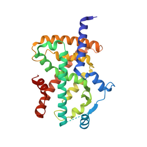 | |
UniProt & NIH Common Fund Data Resources | |||||
PHAROS: P37231 GTEx: ENSG00000132170 | |||||
Entity Groups | |||||
| Sequence Clusters | 30% Identity50% Identity70% Identity90% Identity95% Identity100% Identity | ||||
| UniProt Group | P37231 | ||||
Sequence AnnotationsExpand | |||||
Reference Sequence | |||||
| Ligands 3 Unique | |||||
|---|---|---|---|---|---|
| ID | Chains | Name / Formula / InChI Key | 2D Diagram | 3D Interactions | |
| P1B Download:Ideal Coordinates CCD File | C [auth A], E [auth B] | (5R)-5-{4-[2-(5-ethylpyridin-2-yl)ethoxy]benzyl}-1,3-thiazolidine-2,4-dione C19 H20 N2 O3 S HYAFETHFCAUJAY-QGZVFWFLSA-N |  | ||
| PE4 Download:Ideal Coordinates CCD File | D [auth A] | 2-{2-[2-(2-{2-[2-(2-ETHOXY-ETHOXY)-ETHOXY]-ETHOXY}-ETHOXY)-ETHOXY]-ETHOXY}-ETHANOL C16 H34 O8 PJWQOENWHPEPKI-UHFFFAOYSA-N |  | ||
| EPE Download:Ideal Coordinates CCD File | F [auth B] | 4-(2-HYDROXYETHYL)-1-PIPERAZINE ETHANESULFONIC ACID C8 H18 N2 O4 S JKMHFZQWWAIEOD-UHFFFAOYSA-N |  | ||
| Length ( Å ) | Angle ( ˚ ) |
|---|---|
| a = 92.861 | α = 90 |
| b = 60.411 | β = 103.01 |
| c = 119.849 | γ = 90 |
| Software Name | Purpose |
|---|---|
| REFMAC | refinement |
| HKL-2000 | data reduction |
| SCALEPACK | data scaling |
| PHASER | phasing |


