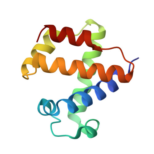
Ligand Migration in the Apolar Tunnel of Cerebratulus Lacteus Mini-Hemoglobin.
Pesce, A., Nardini, M., Dewilde, S., Capece, L., Marti, M.A., Congia, S., Salter, M.D., Blouin, G.C., Estrin, D.A., Ascenzi, P., Moens, L., Bolognesi, M., Olson, J.S.(2011) J Biological Chem 286: 5347
- PubMed: 21147768 Search on PubMedSearch on PubMed Central
- DOI: https://doi.org/10.1074/jbc.M110.169045
- Primary Citation Related Structures:
2XKG, 2XKH, 2XKI - PubMed Abstract:
The large apolar tunnel traversing the mini-hemoglobin from Cerebratulus lacteus (CerHb) has been examined by x-ray crystallography, ligand binding kinetics, and molecular dynamic simulations. The addition of 10 atm of xenon causes loss of diffraction in wild-type (wt) CerHbO(2) crystals, but Leu-86(G12)Ala CerHbO(2), which has an increased tunnel volume, stably accommodates two discrete xenon atoms: one adjacent to Leu-86(G12) and another near Ala-55(E18). Molecular dynamics simulations of ligand migration in wt CerHb show a low energy pathway through the apolar tunnel when Leu or Ala, but not Phe or Trp, is present at the 86(G12) position. The addition of 10-15 atm of xenon to solutions of wt CerHbCO and L86A CerHbCO causes 2-3-fold increases in the fraction of geminate ligand recombination, indicating that the bound xenon blocks CO escape. This idea was confirmed by L86F and L86W mutations, which cause even larger increases in the fraction of geminate CO rebinding, 2-5-fold decreases in the bimolecular rate constants for ligand entry, and large increases in the computed energy barriers for ligand movement through the apolar tunnel. Both the addition of xenon to the L86A mutant and oxidation of wt CerHb heme iron cause the appearance of an out Gln-44(E7) conformer, in which the amide side chain points out toward the solvent and appears to lower the barrier for ligand escape through the E7 gate. However, the observed kinetics suggest little entry and escape (≤ 25%) through the E7 pathway, presumably because the in Gln-44(E7) conformer is thermodynamically favored.
- Department of Physics, University of Genova, Via Dodecaneso 33, 16146 Genova, Italy.
Organizational Affiliation: