2XH1 | pdb_00002xh1

Crystal structure of human KAT II-inhibitor complex


Experimental Data Snapshot

  • Method: X-RAY DIFFRACTION
  • Resolution: 2.10 Å
  • R-Value Free: 
    0.241 (Depositor), 0.238 (DCC) 
  • R-Value Work: 
    0.188 (Depositor), 0.191 (DCC) 
  • R-Value Observed: 
    0.189 (Depositor) 

Starting Model: experimental
View more details

wwPDB Validation 3D Report Full Report

Validation slider image for 2XH1

Ligand Structure Quality Assessment 


This is version 1.2 of the entry. See complete history

Literature

Crystal Structure-Based Selective Targeting of the Pyridoxal 5'-Phsosphate Dependent Enzyme Kynurenine Aminotransferase II for Cognitive Enhancement

Rossi, F.Casazza, V.Garavaglia, S.Sathyasaikumar, K.V.Schwarcz, R.Kojima, S.I.Okuwaki, K.Ono, S.I.Kajii, Y.Rizzi, M.

(2010) J Med Chem 53: 5684

  • DOI: https://doi.org/10.1021/jm100464k
  • Primary Citation Related Structures: 
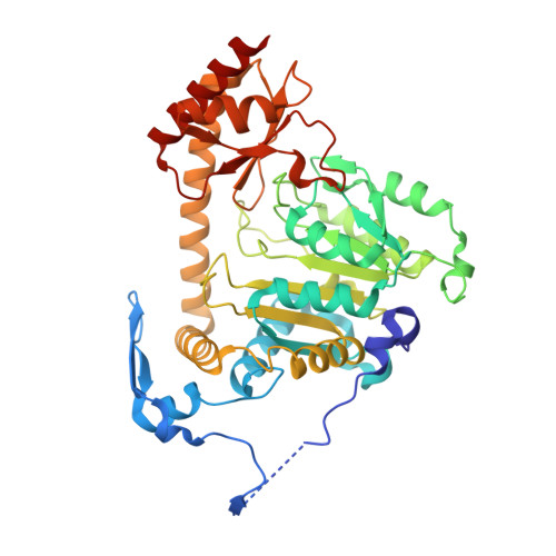
    2XH1

  • PubMed Abstract: 

    Fluctuations in the brain levels of the neuromodulator kynurenic acid may control cognitive processes and play a causative role in several catastrophic brain diseases. Elimination of the pyridoxal 5'-phosphate dependent enzyme kynurenine aminotransferase II reduces cerebral kynurenic acid synthesis and has procognitive effects. The present description of the crystal structure of human kynurenine aminotransferase II in complex with its potent and specific primary amine-bearing fluoroquinolone inhibitor (S)-(-)-9-(4-aminopiperazin-1-yl)-8-fluoro-3-methyl-6-oxo-2,3-dihydro-6H-1-oxa-3a-azaphenalene-5-carboxylic acid (BFF-122) should facilitate the structure-based development of cognition-enhancing drugs. From a medicinal chemistry perspective our results demonstrate that the issue of inhibitor specificity for highly conserved PLP-dependent enzymes could be successfully addressed.


  • Organizational Affiliation
    • University of Piemonte Orientale Amedeo Avogadro, Novara, Italy.

Macromolecule Content 

  • Total Structure Weight: 96.23 kDa 
  • Atom Count: 6,972 
  • Modeled Residue Count: 824 
  • Deposited Residue Count: 850 
  • Unique protein chains: 1

Macromolecules

Find similar proteins by:|  3D Structure
Entity ID: 1
MoleculeChains  Sequence LengthOrganismDetailsImage
KYNURENINE/ALPHA-AMINOADIPATE AMINOTRANSFERASE, MITOCHONDRIAL
A, B
425Homo sapiensMutation(s): 1 
EC: 2.6.1.7 (PDB Primary Data), 2.6.1.39 (PDB Primary Data), 2.6.1.4 (UniProt), 2.6.1.63 (UniProt), 2.6.1.73 (UniProt)
UniProt & NIH Common Fund Data Resources
Find proteins for Q8N5Z0 (Homo sapiens)
Explore Q8N5Z0 
Go to UniProtKB:  Q8N5Z0
PHAROS:  Q8N5Z0
GTEx:  ENSG00000109576 
Entity Groups
Sequence Clusters30% Identity50% Identity70% Identity90% Identity95% Identity100% Identity
UniProt GroupQ8N5Z0
Sequence Annotations
Expand
Reference Sequence

Small Molecules

Ligands 4 Unique
IDChains Name / Formula / InChI Key2D Diagram3D Interactions
BF5

Query on BF5


Download:Ideal Coordinates CCD File
C [auth A],
F [auth B]
(3S)-10-(4-AMINOPIPERAZIN-1-YL)-9-FLUORO-7-HYDROXY-3-METHYL-2,3-DIHYDRO-8H-[1,4]OXAZINO[2,3,4-IJ]QUINOLINE-6-CARBOXYLATE
C17 H19 F N4 O4
XSSSNYCVNIDBIB-VIFPVBQESA-M
PLP

Query on PLP


Download:Ideal Coordinates CCD File
D [auth A],
G [auth B]
PYRIDOXAL-5'-PHOSPHATE
C8 H10 N O6 P
NGVDGCNFYWLIFO-UHFFFAOYSA-N
IOD

Query on IOD


Download:Ideal Coordinates CCD File
E [auth A]IODIDE ION
I
XMBWDFGMSWQBCA-UHFFFAOYSA-M
GOL

Query on GOL


Download:Ideal Coordinates CCD File
H [auth B]GLYCEROL
C3 H8 O3
PEDCQBHIVMGVHV-UHFFFAOYSA-N

Experimental Data & Validation

Experimental Data

  • Method: X-RAY DIFFRACTION
  • Resolution: 2.10 Å
  • R-Value Free:  0.241 (Depositor), 0.238 (DCC) 
  • R-Value Work:  0.188 (Depositor), 0.191 (DCC) 
  • R-Value Observed: 0.189 (Depositor) 
Space Group: P 21 21 2
Unit Cell:
Length ( Å )Angle ( ˚ )
a = 98.323α = 90
b = 152.857β = 90
c = 60.8γ = 90
Software Package:
Software NamePurpose
REFMACrefinement
MOSFLMdata reduction
SCALAdata scaling
AMoREphasing

Structure Validation

View Full Validation Report


Ligand Structure Quality Assessment 


Entry History 

Deposition Data

Revision History  (Full details and data files)

  • Version 1.0: 2010-07-28
    Type: Initial release
  • Version 1.1: 2011-10-26
    Changes: Database references, Version format compliance
  • Version 1.2: 2023-12-20
    Changes: Data collection, Database references, Derived calculations, Other, Refinement description