2XGY | pdb_00002xgy

Complex of Rabbit Endogenous Lentivirus (RELIK)Capsid with Cyclophilin A


Experimental Data Snapshot

  • Method: X-RAY DIFFRACTION
  • Resolution: 1.80 Å
  • R-Value Free: 
    0.187 (Depositor), 0.188 (DCC) 
  • R-Value Work: 
    0.156 (Depositor) 
  • R-Value Observed: 
    0.157 (Depositor) 

wwPDB Validation 3D Report Full Report

Validation slider image for 2XGY

This is version 1.3 of the entry. See complete history

Literature

Structural and Functional Analysis of Prehistoric Lentiviruses Uncovers an Ancient Molecular Interface.

Goldstone, D.C.Yap, M.W.Robertson, L.E.Haire, L.F.Taylor, W.R.Katzourakis, A.Stoye, J.P.Taylor, I.A.

(2010) Cell Host Microbe 8: 248

  • DOI: https://doi.org/10.1016/j.chom.2010.08.006
  • Primary Citation Related Structures: 
    2XGU, 2XGV, 2XGY

  • PubMed Abstract: 

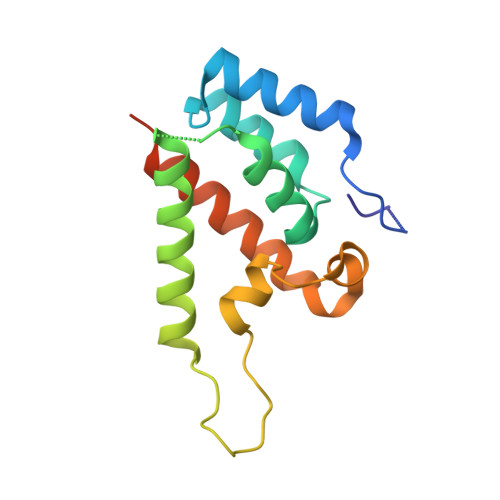
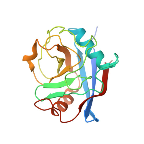
    Lentiviruses are widespread in a variety of vertebrates, often associated with chronic disease states. However, until the recent discovery of the prehistoric endogenous lentiviruses in rabbits (RELIK) and lemurs (PSIV), it was thought that lentiviruses had no capacity for germline integration and were only spread horizontally in an exogenous fashion. The existence of RELIK and PSIV refuted these ideas, revealing lentiviruses to be present in a range of mammals, capable of germline integration, and far more ancient than previously thought. Using Gag sequences reconstructed from the remnants of these prehistoric lentiviruses, we have produced chimeric lentiviruses capable of infecting nondividing cells and determined structures of capsid domains from PSIV and RELIK. We show that the structures from these diverse viruses are highly similar, containing features found in modern-day lentiviruses, including a functional cyclophilin-binding loop. Together, these data provide evidence for an ancient capsid-cyclophilin interaction preserved throughout lentiviral evolution.


  • Organizational Affiliation
    • Division of Molecular Structure, MRC National Institute for Medical Research, The Ridgeway, Mill Hill, London, UK.

Macromolecule Content 

  • Total Structure Weight: 35.52 kDa 
  • Atom Count: 2,616 
  • Modeled Residue Count: 294 
  • Deposited Residue Count: 322 
  • Unique protein chains: 2

Macromolecules

Find similar proteins by:|  3D Structure
Entity ID: 1
MoleculeChains  Sequence LengthOrganismDetailsImage
RELIK CAPSID N-TERMINAL DOMAIN149Oryctolagus cuniculusMutation(s): 0 
Find similar proteins by:|  3D Structure
Entity ID: 2
MoleculeChains  Sequence LengthOrganismDetailsImage
PEPTIDYL-PROLYL CIS-TRANS ISOMERASE A173Homo sapiensMutation(s): 0 
EC: 5.2.1.8
UniProt & NIH Common Fund Data Resources
Find proteins for P62937 (Homo sapiens)
Explore P62937 
Go to UniProtKB:  P62937
PHAROS:  P62937
GTEx:  ENSG00000196262 
Entity Groups
Sequence Clusters30% Identity50% Identity70% Identity90% Identity95% Identity100% Identity
UniProt GroupP62937
Sequence Annotations
Expand
Reference Sequence

Experimental Data & Validation

Experimental Data

  • Method: X-RAY DIFFRACTION
  • Resolution: 1.80 Å
  • R-Value Free:  0.187 (Depositor), 0.188 (DCC) 
  • R-Value Work:  0.156 (Depositor) 
  • R-Value Observed: 0.157 (Depositor) 
Space Group: P 61
Unit Cell:
Length ( Å )Angle ( ˚ )
a = 117.158α = 90
b = 117.158β = 90
c = 49.655γ = 120
Software Package:
Software NamePurpose
PHENIXrefinement
DENZOdata reduction
SCALEPACKdata scaling
PHASERphasing

Structure Validation

View Full Validation Report


Entry History 

Deposition Data

Revision History  (Full details and data files)

  • Version 1.0: 2010-09-22
    Type: Initial release
  • Version 1.1: 2011-05-08
    Changes: Version format compliance
  • Version 1.2: 2011-07-13
    Changes: Version format compliance
  • Version 1.3: 2024-05-08
    Changes: Data collection, Database references, Derived calculations, Other