2XGA | pdb_00002xga

MTSL spin-labelled Shigella Flexneri Spa15


Experimental Data Snapshot

  • Method: X-RAY DIFFRACTION
  • Resolution: 2.30 Å
  • R-Value Free: 
    0.264 (Depositor), 0.257 (DCC) 
  • R-Value Work: 
    0.224 (Depositor), 0.221 (DCC) 
  • R-Value Observed: 
    0.226 (Depositor) 

Starting Model: experimental
View more details

wwPDB Validation 3D Report Full Report

Validation slider image for 2XGA

Ligand Structure Quality Assessment 


This is version 1.4 of the entry. See complete history

Literature

Shigella Flexneri Spa15 Crystal Structure Verified in Solution by Double Electron Electron Resonance.

Lillington, J.E.D.Lovett, J.E.Johnson, S.Roversi, P.Timmel, C.R.Lea, S.M.

(2011) J Mol Biology 405: 427

  • DOI: https://doi.org/10.1016/j.jmb.2010.10.053
  • Primary Citation Related Structures: 
    2XGA

  • PubMed Abstract: 

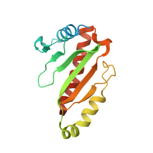
    Shigella flexneri Spa15 is a chaperone of the type 3 secretion system, which binds a number of effectors to ensure their stabilization prior to secretion. One of these effectors is IpgB1, a mimic of the human Ras-like Rho guanosine triphosphatase RhoG. In this study, Spa15 alone and in complex with IpgB1 has been studied by double electron electron resonance, an experiment that gives distance information showing the spacial separation of attached spin labels. This distance is explained by determining the crystal structure of the spin-labeled Spa15 where labels are seen to be buried in hydrophobic pockets. The double electron electron resonance experiment on the Spa15 complex with IpgB1 shows that IpgB1 does not bind Spa15 in the same way as is seen in the homologous Salmonella sp. chaperone:effector complex InvB:SipA.


  • Organizational Affiliation
    • Inorganic Chemistry Laboratory, University of Oxford, OX1 3QR, UK.

Macromolecule Content 

  • Total Structure Weight: 35.12 kDa 
  • Atom Count: 2,156 
  • Modeled Residue Count: 255 
  • Deposited Residue Count: 306 
  • Unique protein chains: 1

Macromolecules

Find similar proteins by:|  3D Structure
Entity ID: 1
MoleculeChains  Sequence LengthOrganismDetailsImage
SURFACE PRESENTATION OF ANTIGENS PROTEIN SPAK
A, B
153Shigella flexneriMutation(s): 0 
UniProt
Find proteins for P35530 (Shigella flexneri)
Explore P35530 
Go to UniProtKB:  P35530
Entity Groups
Sequence Clusters30% Identity50% Identity70% Identity90% Identity95% Identity100% Identity
UniProt GroupP35530
Sequence Annotations
Expand
Reference Sequence

Small Molecules

Ligands 1 Unique
IDChains Name / Formula / InChI Key2D Diagram3D Interactions
MTN

Query on MTN


Download:Ideal Coordinates CCD File
C [auth A],
D [auth B]
S-[(1-oxyl-2,2,5,5-tetramethyl-2,5-dihydro-1H-pyrrol-3-yl)methyl] methanesulfonothioate
C10 H18 N O3 S2
MXZPGYFBZHBAQM-UHFFFAOYSA-N

Experimental Data & Validation

Experimental Data

  • Method: X-RAY DIFFRACTION
  • Resolution: 2.30 Å
  • R-Value Free:  0.264 (Depositor), 0.257 (DCC) 
  • R-Value Work:  0.224 (Depositor), 0.221 (DCC) 
  • R-Value Observed: 0.226 (Depositor) 
Space Group: P 1 21 1
Unit Cell:
Length ( Å )Angle ( ˚ )
a = 55.98α = 90
b = 50.38β = 117.03
c = 57.68γ = 90
Software Package:
Software NamePurpose
REFMACrefinement
MOSFLMdata reduction
SCALAdata scaling
MOLREPphasing

Structure Validation

View Full Validation Report


Ligand Structure Quality Assessment 


Entry History 

Deposition Data

Revision History  (Full details and data files)

  • Version 1.0: 2010-12-01
    Type: Initial release
  • Version 1.1: 2011-05-12
    Changes: Version format compliance
  • Version 1.2: 2011-07-13
    Changes: Version format compliance
  • Version 1.3: 2023-12-20
    Changes: Advisory, Data collection, Database references, Derived calculations, Other, Refinement description
  • Version 1.4: 2024-11-20
    Changes: Structure summary