2XDY

Crystal structure of the N. crassa QDE-2 AGO MID domain


Experimental Data Snapshot

  • Method: X-RAY DIFFRACTION
  • Resolution: 2.20 Å
  • R-Value Free: 0.223 
  • R-Value Work: 0.193 
  • R-Value Observed: 0.194 

wwPDB Validation   3D Report Full Report


This is version 1.2 of the entry. See complete history


Literature

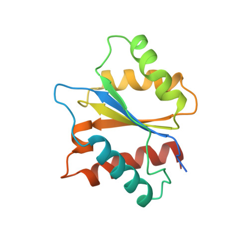
Crystal Structure and Ligand Binding of the Mid Domain of a Eukaryotic Argonaute Protein.

Boland, A.Tritschler, F.Heimstaedt, S.Izaurralde, E.Weichenrieder, O.

(2010) EMBO Rep 11: 522

  • DOI: https://doi.org/10.1038/embor.2010.81
  • Primary Citation of Related Structures:  
    2XDY

  • PubMed Abstract: 

    Argonaute (AGO) proteins are core components of RNA-induced silencing complexes and have essential roles in RNA-mediated gene silencing. They are characterized by a bilobal architecture, consisting of one lobe containing the amino-terminal and PAZ domains and another containing the MID and PIWI domains. Except for the PAZ domain, structural information on eukaryotic AGO domains is not yet available. In this study, we report the crystal structure of the MID domain of the eukaryotic AGO protein QDE-2 from Neurospora crassa. This domain adopts a Rossmann-like fold and recognizes the 5'-terminal nucleotide of a guide RNA in a manner similar to its prokaryotic counterparts. The 5'-nucleotide-binding site shares common residues with a second, adjacent ligand-binding site, suggesting a mechanism for the cooperative binding of ligands to the MID domain of eukaryotic AGOs.


  • Organizational Affiliation

    Department of Biochemistry, Max Planck Institute for Developmental Biology, Spemannstrasse 35, Tübingen D-72076, Germany.


Macromolecules
Find similar proteins by:  (by identity cutoff)  |  3D Structure
Entity ID: 1
MoleculeChains Sequence LengthOrganismDetailsImage
POST-TRANSCRIPTIONAL GENE SILENCING PROTEIN QDE-2145Neurospora crassaMutation(s): 0 
UniProt
Find proteins for Q9P8T1 (Neurospora crassa)
Explore Q9P8T1 
Go to UniProtKB:  Q9P8T1
Entity Groups  
Sequence Clusters30% Identity50% Identity70% Identity90% Identity95% Identity100% Identity
UniProt GroupQ9P8T1
Sequence Annotations
Expand
  • Reference Sequence
Experimental Data & Validation

Experimental Data

  • Method: X-RAY DIFFRACTION
  • Resolution: 2.20 Å
  • R-Value Free: 0.223 
  • R-Value Work: 0.193 
  • R-Value Observed: 0.194 
  • Space Group: C 2 2 21
Unit Cell:
Length ( Å )Angle ( ˚ )
a = 80.1α = 90
b = 91.39β = 90
c = 42.93γ = 90
Software Package:
Software NamePurpose
XDSdata reduction
XSCALEdata scaling
SHELXphasing
autoSHARPphasing
PHENIXrefinement

Structure Validation

View Full Validation Report


Entry History 

Deposition Data

Revision History  (Full details and data files)

  • Version 1.0: 2010-06-16
    Type: Initial release
  • Version 1.1: 2011-05-19
    Changes: Version format compliance
  • Version 1.2: 2011-07-13
    Changes: Version format compliance