2XBD | pdb_00002xbd

INTERNAL XYLAN BINDING DOMAIN FROM CELLULOMONAS FIMI XYLANASE D, NMR, MINIMIZED AVERAGE STRUCTURE


Experimental Data Snapshot

  • Method: SOLUTION NMR
  • Conformers Calculated: 50 
  • Conformers Submitted: 
  • Selection Criteria: MINIMIZED AVERAGE STRUCTURE, CALCULATED FROM 38 LOWEST ENERGY STRUCTURES 

wwPDB Validation 3D Report Full Report

Validation slider image for 2XBD

This is version 1.4 of the entry. See complete history

Literature

A family IIb xylan-binding domain has a similar secondary structure to a homologous family IIa cellulose-binding domain but different ligand specificity.

Simpson, P.J.Bolam, D.N.Cooper, A.Ciruela, A.Hazlewood, G.P.Gilbert, H.J.Williamson, M.P.

(1999) Structure 7: 853-864

  • DOI: https://doi.org/10.1016/s0969-2126(99)80108-7
  • Primary Citation Related Structures: 
    1XBD, 2XBD

  • PubMed Abstract: 

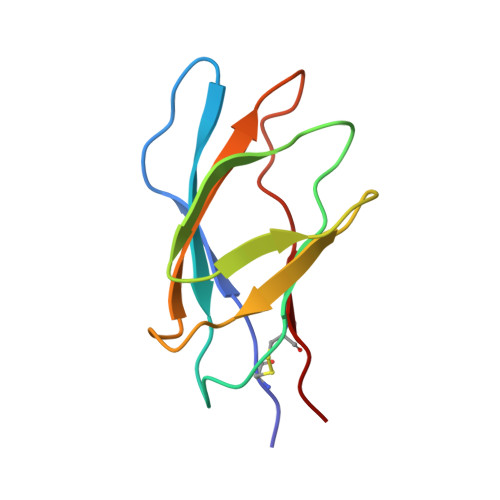
    Many enzymes that digest polysaccharides contain separate polysaccharide-binding domains. Structures have been previously determined for a number of cellulose-binding domains (CBDs) from cellulases. The family IIb xylan-binding domain 1 (XBD1) from Cellulomonas fimi xylanase D is shown to bind xylan but not cellulose. Its structure is similar to that of the homologous family IIa CBD from C. fimi Cex, consisting of two four-stranded beta sheets that form a twisted 'beta sandwich'. The xylan-binding site is a groove made from two tryptophan residues that stack against the faces of the sugar rings, plus several hydrogen-bonding polar residues. The biggest difference between the family IIa and IIb domains is that in the former the solvent-exposed tryptophan sidechains are coplanar, whereas in the latter they are perpendicular, forming a twisted binding site. The binding sites are therefore complementary to the secondary structures of the ligands cellulose and xylan. XBD1 and CexCBD represent a striking example of two proteins that have high sequence similarity but a different function.


  • Organizational Affiliation
    • Department of Molecular Biology and Biotechnology, Krebs Institute, University of Sheffield, UK.

Macromolecule Content 

  • Total Structure Weight: 8.79 kDa 
  • Atom Count: 615 
  • Modeled Residue Count: 87 
  • Deposited Residue Count: 87 
  • Unique protein chains: 1

Macromolecules

Find similar proteins by:|  3D Structure
Entity ID: 1
MoleculeChains  Sequence LengthOrganismDetailsImage
XYLANASE D87Cellulomonas fimiMutation(s): 0 
EC: 3.2.1.8 (PDB Primary Data), 3.5.1 (UniProt)
UniProt
Find proteins for P54865 (Cellulomonas fimi)
Explore P54865 
Go to UniProtKB:  P54865
Entity Groups
Sequence Clusters30% Identity50% Identity70% Identity90% Identity95% Identity100% Identity
UniProt GroupP54865
Sequence Annotations
Expand
Reference Sequence

Experimental Data & Validation

Experimental Data

  • Method: SOLUTION NMR
  • Conformers Calculated: 50 
  • Conformers Submitted: 
  • Selection Criteria: MINIMIZED AVERAGE STRUCTURE, CALCULATED FROM 38 LOWEST ENERGY STRUCTURES 

Structure Validation

View Full Validation Report


Entry History 

Deposition Data

Revision History  (Full details and data files)

  • Version 1.0: 1999-07-21
    Type: Initial release
  • Version 1.1: 2008-03-25
    Changes: Version format compliance
  • Version 1.2: 2011-07-13
    Changes: Version format compliance
  • Version 1.3: 2022-03-16
    Changes: Database references, Derived calculations, Other
  • Version 1.4: 2024-10-30
    Changes: Data collection, Structure summary