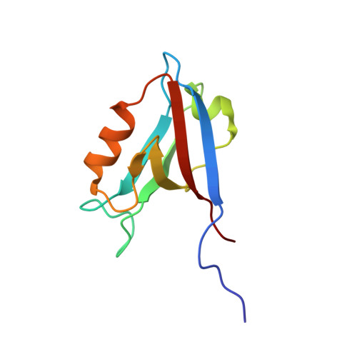
2X7Z

Crystal Structure of the SAP97 PDZ2 I342W C378A mutant protein domain


Experimental Data Snapshot

  • Method: X-RAY DIFFRACTION
  • Resolution: 2.00 Å
  • R-Value Free: 0.232 
  • R-Value Work: 0.194 
  • R-Value Observed: 0.196 

Starting Model: experimental
View more details

wwPDB Validation   3D Report Full Report


This is version 1.4 of the entry. See complete history


Literature

The Plastic Energy Landscape of Protein Folding: A Triangular Folding Mechanism with an Equilibrium Intermediate for a Small Protein Domain.

Haq, S.R.Jurgens, M.C.Chi, C.N.Elfstrom, L.Koh, C.S.Selmer, M.Gianni, S.Jemth, P.

(2010) J Biol Chem 285: 18051

  • DOI: https://doi.org/10.1074/jbc.M110.110833
  • Primary Citation of Related Structures:  
    2X7Z

  • PubMed Abstract: 

    Protein domains usually fold without or with only transiently populated intermediates, possibly to avoid misfolding, which could result in amyloidogenic disease. Whether observed intermediates are productive and obligatory species on the folding reaction pathway or dispensable by-products is a matter of debate. Here, we solved the crystal structure of a small protein domain, SAP97 PDZ2 I342W C378A, and determined its folding pathway. The presence of a folding intermediate was demonstrated both by single and double-mixing kinetic experiments using urea-induced (un)folding as well as ligand-induced folding. This protein domain was found to fold via a triangular scheme, where the folding intermediate could be either on- or off-pathway, depending on the experimental conditions. Furthermore, we found that the intermediate was present at equilibrium, which is rarely seen in folding reactions of small protein domains. The folding mechanism observed here illustrates the roughness and plasticity of the protein folding energy landscape, where several routes may be employed to reach the native state. The results also reconcile the folding mechanisms of topological variants within the PDZ domain family.


  • Organizational Affiliation

    Department of Medical Biochemistry and Microbiology, Uppsala University, SE-75123 Uppsala, Sweden.


Macromolecules
Find similar proteins by:  (by identity cutoff)  |  3D Structure
Entity ID: 1
MoleculeChains Sequence LengthOrganismDetailsImage
DISKS LARGE HOMOLOG 199Homo sapiensMutation(s): 2 
UniProt & NIH Common Fund Data Resources
Find proteins for Q12959 (Homo sapiens)
Explore Q12959 
Go to UniProtKB:  Q12959
PHAROS:  Q12959
GTEx:  ENSG00000075711 
Entity Groups  
Sequence Clusters30% Identity50% Identity70% Identity90% Identity95% Identity100% Identity
UniProt GroupQ12959
Sequence Annotations
Expand
  • Reference Sequence
Experimental Data & Validation

Experimental Data

  • Method: X-RAY DIFFRACTION
  • Resolution: 2.00 Å
  • R-Value Free: 0.232 
  • R-Value Work: 0.194 
  • R-Value Observed: 0.196 
  • Space Group: P 43 2 2
Unit Cell:
Length ( Å )Angle ( ˚ )
a = 47.905α = 90
b = 47.905β = 90
c = 123.287γ = 90
Software Package:
Software NamePurpose
REFMACrefinement
XDSdata reduction
XSCALEdata scaling
PHASERphasing

Structure Validation

View Full Validation Report


Entry History 

Deposition Data

Revision History  (Full details and data files)

  • Version 1.0: 2010-03-31
    Type: Initial release
  • Version 1.1: 2011-07-13
    Changes: Advisory, Version format compliance
  • Version 1.2: 2018-01-17
    Changes: Data collection
  • Version 1.3: 2019-03-06
    Changes: Data collection, Experimental preparation
  • Version 1.4: 2023-12-20
    Changes: Data collection, Database references, Derived calculations, Other, Refinement description