2X26

Crystal structure of the periplasmic aliphatic sulphonate binding protein SsuA from Escherichia coli


Experimental Data Snapshot

  • Method: X-RAY DIFFRACTION
  • Resolution: 1.75 Å
  • R-Value Free: 0.225 
  • R-Value Work: 0.186 
  • R-Value Observed: 0.188 

wwPDB Validation   3D Report Full Report


This is version 1.2 of the entry. See complete history


Literature

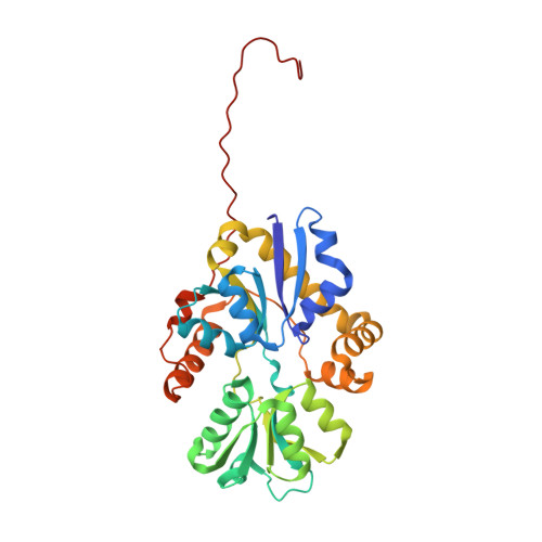
Structure of the Aliphatic Sulfonate-Binding Protein Ssua from Escherichia Coli

Beale, J.Lee, S.Y.Iwata, S.Beis, K.

(2010) Acta Crystallogr Sect F Struct Biol Cryst Commun 66: 391

  • DOI: 10.1107/S1744309110006226
  • Primary Citation of Related Structures:  
    2X26

  • PubMed Abstract: 
  • Sulfur is an essential component for the biosynthesis of the sulfur-containing amino acids L-methionine and L-cysteine. Under sulfur-starvation conditions, bacteria are capable of scavenging sulfur from sulfur-containing compounds and transporting it across membranes ...

    Sulfur is an essential component for the biosynthesis of the sulfur-containing amino acids L-methionine and L-cysteine. Under sulfur-starvation conditions, bacteria are capable of scavenging sulfur from sulfur-containing compounds and transporting it across membranes. Here, the crystal structure of the periplasmic aliphatic sulfonate-binding protein SsuA from Escherichia coli is reported at 1.75 A resolution in the substrate-free state. The overall structure of SsuA resembles the structures of other periplasmic binding proteins and contains two globular domains that form a cleft. Comparison with other periplasmic binding proteins revealed that one of the domains has been displaced by a rigid movement of 17 degrees . Interestingly, the tight crystal packing appears to be mediated by a 13-amino-acid tail from the cloning that folds within the cleft of the next monomer.


    Organizational Affiliation

    Membrane Protein Laboratory, Diamond Light Source, Harwell Science and Innovation Campus, Oxfordshire OX11 0DE, England.


Macromolecules
Find similar proteins by:  (by identity cutoff)  |  3D Structure
Entity ID: 1
MoleculeChainsSequence LengthOrganismDetailsImage
PERIPLASMIC ALIPHATIC SULPHONATES-BINDING PROTEINA, B308Escherichia coli K-12Mutation(s): 0 
Gene Names: ssuAycbOb0936JW0919
UniProt
Find proteins for P75853 (Escherichia coli (strain K12))
Explore P75853 
Go to UniProtKB:  P75853
Entity Groups  
Sequence Clusters30% Identity50% Identity70% Identity90% Identity95% Identity100% Identity
UniProt GroupP75853
Protein Feature View
Expand
  • Reference Sequence
Small Molecules
Ligands 1 Unique
IDChainsName / Formula / InChI Key2D Diagram3D Interactions
GOL
Query on GOL

Download Ideal Coordinates CCD File 
C [auth A],
D [auth B]
GLYCEROL
C3 H8 O3
PEDCQBHIVMGVHV-UHFFFAOYSA-N
 Ligand Interaction
Experimental Data & Validation

Experimental Data

  • Method: X-RAY DIFFRACTION
  • Resolution: 1.75 Å
  • R-Value Free: 0.225 
  • R-Value Work: 0.186 
  • R-Value Observed: 0.188 
  • Space Group: P 21 21 21
Unit Cell:
Length ( Å )Angle ( ˚ )
a = 40.891α = 90
b = 96.026β = 90
c = 142.401γ = 90
Software Package:
Software NamePurpose
XDSdata reduction
SCALAdata scaling
BALBESphasing
MOLREPphasing
REFMACrefinement

Structure Validation

View Full Validation Report


Entry History 

Deposition Data

Revision History  (Full details and data files)

  • Version 1.0: 2010-04-14
    Type: Initial release
  • Version 1.1: 2011-07-13
    Changes: Advisory, Version format compliance
  • Version 1.2: 2019-03-06
    Changes: Data collection, Experimental preparation, Other