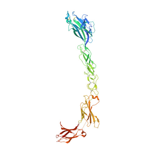
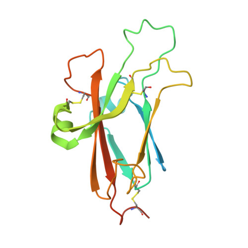
2X11

Crystal structure of the complete EphA2 ectodomain in complex with ephrin A5 receptor binding domain


Experimental Data Snapshot

  • Method: X-RAY DIFFRACTION
  • Resolution: 4.83 Å
  • R-Value Free: 0.314 
  • R-Value Work: 0.312 
  • R-Value Observed: 0.313 

Starting Models: experimental
View more details

wwPDB Validation   3D Report Full Report


This is version 1.4 of the entry. See complete history


Literature

An Extracellular Steric Seeding Mechanism for Eph-Ephrin Signalling Platform Assembly

Seiradake, E.Harlos, K.Sutton, G.Aricescu, A.R.Jones, E.Y.

(2010) Nat Struct Mol Biol 17: 398

  • DOI: https://doi.org/10.1038/nsmb.1782
  • Primary Citation of Related Structures:  
    2X10, 2X11

  • PubMed Abstract: 

    Erythropoetin-producing hepatoma (Eph) receptors are cell-surface protein tyrosine kinases mediating cell-cell communication. Upon activation, they form signaling clusters. We report crystal structures of the full ectodomain of human EphA2 (eEphA2) both alone and in complex with the receptor-binding domain of the ligand ephrinA5 (ephrinA5 RBD). Unliganded eEphA2 forms linear arrays of staggered parallel receptors involving two patches of residues conserved across A-class Ephs. eEphA2-ephrinA5 RBD forms a more elaborate assembly, whose interfaces include the same conserved regions on eEphA2, but rearranged to accommodate ephrinA5 RBD. Cell-surface expression of mutant EphA2s showed that these interfaces are critical for localization at cell-cell contacts and activation-dependent degradation. Our results suggest a 'nucleation' mechanism whereby a limited number of ligand-receptor interactions 'seed' an arrangement of receptors which can propagate into extended signaling arrays.


  • Organizational Affiliation

    Division of Structural Biology, Wellcome Trust Centre for Human Genetics, University of Oxford, Oxford, UK.


Macromolecules
Find similar proteins by:  (by identity cutoff)  |  3D Structure
Entity ID: 1
MoleculeChains Sequence LengthOrganismDetailsImage
EPHRIN TYPE-A RECEPTOR 2545Homo sapiensMutation(s): 0 
EC: 2.7.10.1
UniProt & NIH Common Fund Data Resources
Find proteins for P29317 (Homo sapiens)
Explore P29317 
Go to UniProtKB:  P29317
PHAROS:  P29317
GTEx:  ENSG00000142627 
Entity Groups  
Sequence Clusters30% Identity50% Identity70% Identity90% Identity95% Identity100% Identity
UniProt GroupP29317
Sequence Annotations
Expand
  • Reference Sequence
Find similar proteins by:  (by identity cutoff)  |  3D Structure
Entity ID: 2
MoleculeChains Sequence LengthOrganismDetailsImage
EPHRIN-A5177Homo sapiensMutation(s): 0 
UniProt & NIH Common Fund Data Resources
Find proteins for P52803 (Homo sapiens)
Explore P52803 
Go to UniProtKB:  P52803
PHAROS:  P52803
GTEx:  ENSG00000184349 
Entity Groups  
Sequence Clusters30% Identity50% Identity70% Identity90% Identity95% Identity100% Identity
UniProt GroupP52803
Sequence Annotations
Expand
  • Reference Sequence
Experimental Data & Validation

Experimental Data

  • Method: X-RAY DIFFRACTION
  • Resolution: 4.83 Å
  • R-Value Free: 0.314 
  • R-Value Work: 0.312 
  • R-Value Observed: 0.313 
  • Space Group: P 2 2 21
Unit Cell:
Length ( Å )Angle ( ˚ )
a = 173.63α = 90
b = 59.63β = 90
c = 112.16γ = 90
Software Package:
Software NamePurpose
PHENIXrefinement
XDSdata reduction
XSCALEdata scaling
PHASERphasing

Structure Validation

View Full Validation Report


Entry History 

Deposition Data

Revision History  (Full details and data files)

  • Version 1.0: 2010-03-16
    Type: Initial release
  • Version 1.1: 2011-08-31
    Changes: Database references, Derived calculations, Refinement description, Version format compliance
  • Version 1.2: 2018-02-28
    Changes: Source and taxonomy
  • Version 1.3: 2023-12-20
    Changes: Data collection, Database references, Other, Refinement description
  • Version 1.4: 2024-11-13
    Changes: Structure summary