2X0Y | pdb_00002x0y

Screening-based discovery of drug-like O-GlcNAcase inhibitor scaffolds


Experimental Data Snapshot

  • Method: X-RAY DIFFRACTION
  • Resolution: 2.25 Å
  • R-Value Free: 
    0.246 (Depositor), 0.241 (DCC) 
  • R-Value Work: 
    0.203 (Depositor), 0.196 (DCC) 
  • R-Value Observed: 
    0.204 (Depositor) 

Starting Model: experimental
View more details

wwPDB Validation 3D Report Full Report

Validation slider image for 2X0Y

Ligand Structure Quality Assessment 


This is version 1.3 of the entry. See complete history

Literature

Screening-Based Discovery of Drug-Like O-Glcnacase Inhibitor Scaffolds

Dorfmueller, H.C.Van Aalten, D.M.F.

(2010) FEBS Lett 584: 694

  • DOI: https://doi.org/10.1016/j.febslet.2009.12.020
  • Primary Citation Related Structures: 
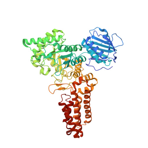
    2X0Y

  • PubMed Abstract: 

    O-GlcNAcylation is an essential posttranslational modification in metazoa. Modulation of O-GlcNAc levels with small molecule inhibitors of O-GlcNAc hydrolase (OGA) is a useful strategy to probe the role of this modification in a range of cellular processes. Here we report the discovery of novel, low molecular weight and drug-like O-GlcNAcase inhibitor scaffolds by high-throughput screening. Kinetic and X-ray crystallographic analyses of the binding modes with human/bacterial O-GlcNAcases identify some of these as competitive inhibitors. Comparative kinetic experiments with the mechanistically related human lysosomal hexosaminidases reveal that three of the inhibitor scaffolds show selectivity towards human OGA. These scaffolds provide attractive starting points for the development of non-carbohydrate, drug-like OGA inhibitors.


  • Organizational Affiliation
    • Division of Molecular Microbiology, College of Life Sciences, University of Dundee, Dundee, Scotland, UK.

Macromolecule Content 

  • Total Structure Weight: 133.89 kDa 
  • Atom Count: 10,123 
  • Modeled Residue Count: 1,169 
  • Deposited Residue Count: 1,188 
  • Unique protein chains: 1

Macromolecules

Find similar proteins by:|  3D Structure
Entity ID: 1
MoleculeChains  Sequence LengthOrganismDetailsImage
O-GLCNACASE NAGJ
A, B
594Clostridium perfringensMutation(s): 0 
EC: 3.2.1.52 (PDB Primary Data), 3.2.1.169 (UniProt)
UniProt
Find proteins for Q0TR53 (Clostridium perfringens (strain ATCC 13124 / DSM 756 / JCM 1290 / NCIMB 6125 / NCTC 8237 / Type A))
Explore Q0TR53 
Go to UniProtKB:  Q0TR53
Entity Groups
Sequence Clusters30% Identity50% Identity70% Identity90% Identity95% Identity100% Identity
UniProt GroupQ0TR53
Sequence Annotations
Expand
Reference Sequence

Small Molecules

Ligands 1 Unique
IDChains Name / Formula / InChI Key2D Diagram3D Interactions
X0T

Query on X0T


Download:Ideal Coordinates CCD File
C [auth A],
D [auth B]
7-[(2S)-2,3-DIHYDROXYPROPYL]-1,3-DIMETHYL-3,7-DIHYDRO-1H-PURINE-2,6-DIONE
C10 H14 N4 O4
KSCFJBIXMNOVSH-LURJTMIESA-N

Experimental Data & Validation

Experimental Data

  • Method: X-RAY DIFFRACTION
  • Resolution: 2.25 Å
  • R-Value Free:  0.246 (Depositor), 0.241 (DCC) 
  • R-Value Work:  0.203 (Depositor), 0.196 (DCC) 
  • R-Value Observed: 0.204 (Depositor) 
Space Group: I 21 21 21
Unit Cell:
Length ( Å )Angle ( ˚ )
a = 130.405α = 90
b = 148.549β = 90
c = 151.97γ = 90
Software Package:
Software NamePurpose
REFMACrefinement
DENZOdata scaling
REFMACphasing

Structure Validation

View Full Validation Report


Ligand Structure Quality Assessment 


Entry History 

Deposition Data

Revision History  (Full details and data files)

  • Version 1.0: 2010-01-12
    Type: Initial release
  • Version 1.1: 2011-06-02
    Changes: Version format compliance
  • Version 1.2: 2011-07-13
    Changes: Version format compliance
  • Version 1.3: 2023-12-20
    Changes: Data collection, Database references, Derived calculations, Other, Refinement description